Figure 1. Mouse model of limbal stem cell
deficiency created using limbus to limbus scraping. Fluorescein
staining pattern of a normal cornea showing minimal staining due to
dryness (A). Immediately after total corneal epithelial
debridement there was a total epithelial defect (B). Three weeks
after total removal of the corneal epithelium, fluorescein staining
revealed diffuse punctate epithelial staining (C). Two months
after total debridement, the cornea has developed progressive
superficial neovascularization (D).
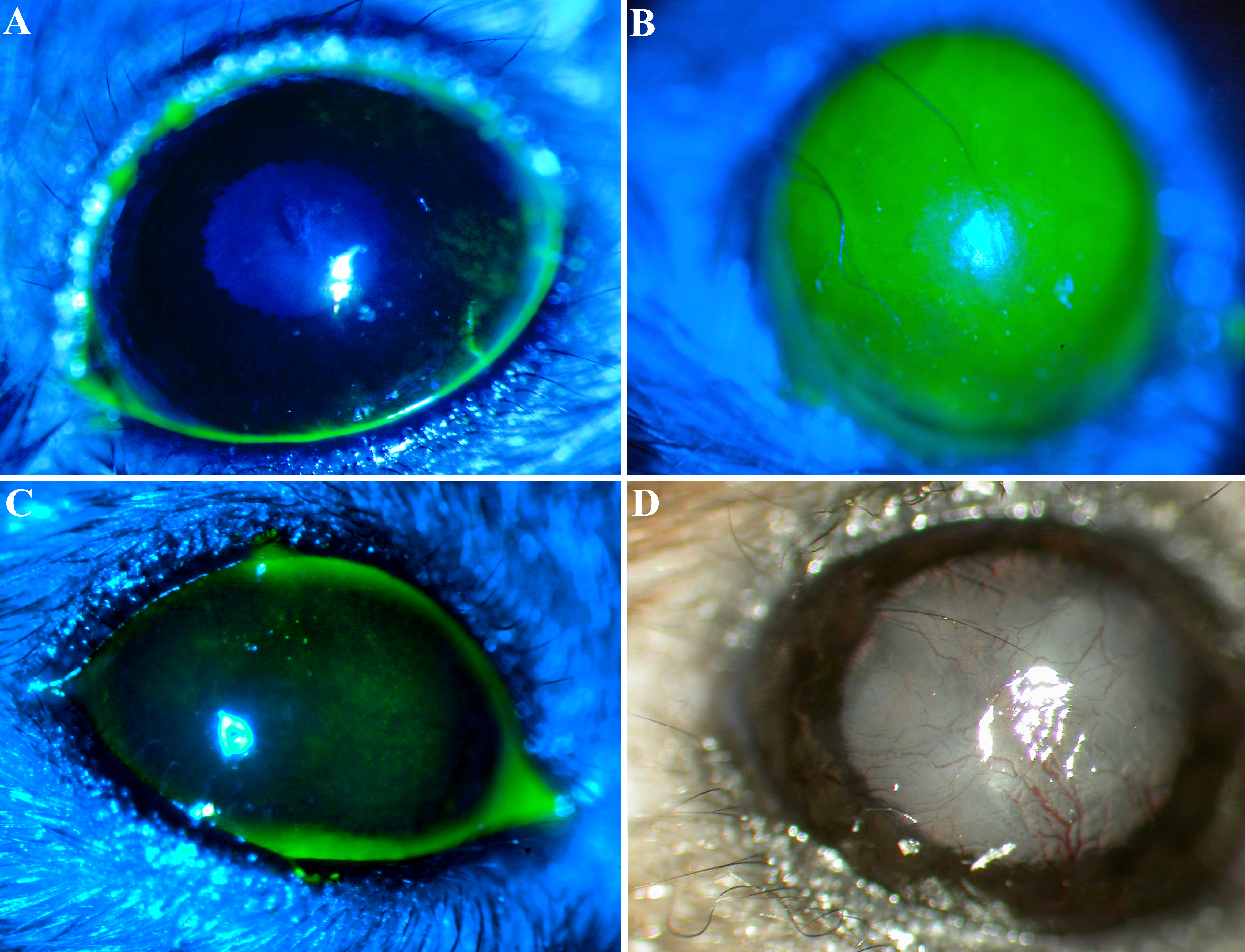
 Figure 1 of Amirjamshidi, Mol Vis 2011; 17:658-666.