Figure 5. Immunofluorescence and confocal
microscopy were used to detect the expression of PAX and F-actin.
F-actin was stained with FITC (A, D, and G) and
PAX was stained with Texas red (B, E, and H).
The areas of co-localization appear yellow in the merged sections (C,
F, and I). When the cells were pretreated with
genistein, the expression of PAX decreased (B, E, and H).
Incubation
with F. solani spores induced alterations in the
F-actin microfilaments of the HCECs (A, D, and G).
These
results showed that the untreated HCECs exhibited normal
morphology, while an actin rearrangement was noted in cells incubated
with the F. solani spores. A combined treatment with genistein
and the spores decreased the polymerization of actin, and the HCECs
became more spreading than in the group incubated with F. solani
spores.
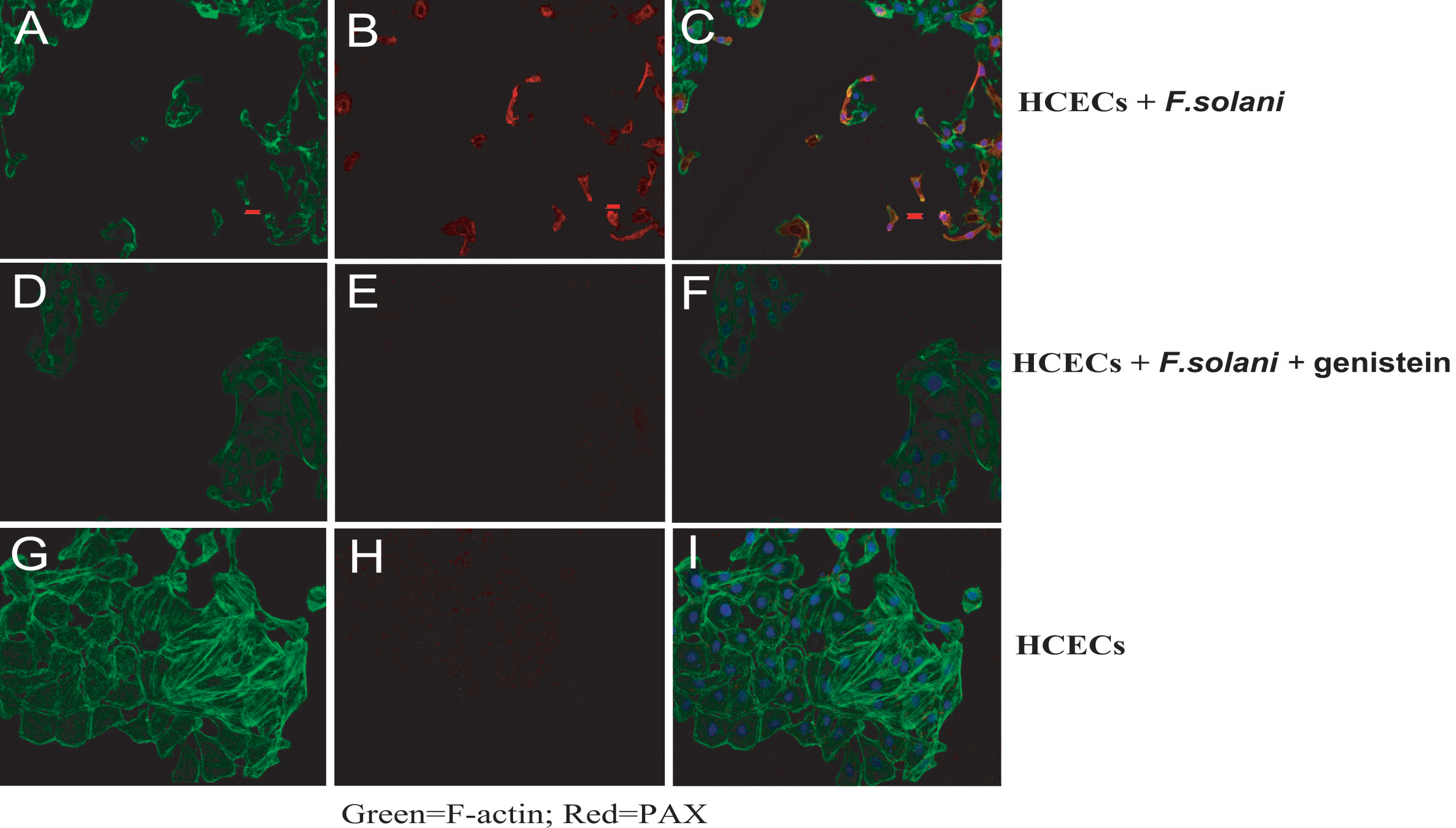
 Figure 5 of Pan, Mol Vis 2011; 17:638-646.