Figure 3. Changes in the ultrastructure after HCECs were coincubated with F. solani spores. A-D: Scanning electron micrographs of HCECs coincubated with F. solani spores. After coincubation for 6 h, the F. solani spores (arrow) began to attach to the surface of the HCECs as seen in panels (A) and (D). The number of adhered cells increased at 8 h after coincubation (B, arrow). Most of the cells presented a great number of spores congregated to the projections of plasma membrane (A-E, arrow). Damage to the HCECs can be seen in panels B, E, C, and F. Ruptured and extensively destroyed membrane with spores adhering to it (C, arrow) and characteristics of dead cells (F, arrow) at 10 h after coincubation. Original magnification: (A, B, and C) 2,000×; (D, E, and F) 800×. G-L: Transmission electron micrographs of cells incubated with HCECs and F. solani spores. Photograph shows F. solani spores attached to the plasma membrane, followed by the subsequent formation of cell projections around it (8 h, G, and J, arrow). Then, spores are internalized into the cell cytoplasm and cell organelles appeared to be destroyed at 10 h (H and I). The ultrastructures of most HCECs show confused organelle structure degeneration of the nucleus (H, arrow), and vacuolization of the mitochondria (K, arrow) at 10 h after incubation. Original magnification: (G) 8,000×; (I and J) 5,000×; (H) 3,000×; (K) 20,000×; (L) 12,000×.
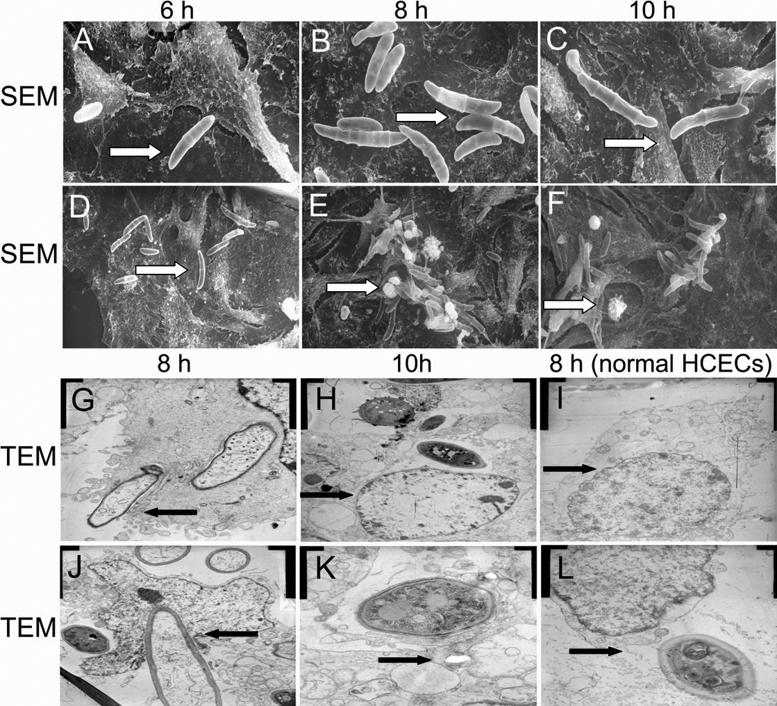
 Figure 3 of
Pan, Mol Vis 2011; 17:638-646.
Figure 3 of
Pan, Mol Vis 2011; 17:638-646.