Figure 4. Cytotoxicity assay on Müller
cell. A: The percentage of trypan blue positive cells (y-axis)
was plotted against the concentration of the test compounds used
(x-axis). The concentration unit was micro molar and the open box
represented a 95% confidence limit. 5-FU at 100 µM that was the highest
concentration tested, caused 20% death of the cell culture, which was
equivalent to HDP-PMEG at 11 µM or DNR at 0.1 µM. B: Exemplary
images from the Müller cell cytotoxicity study: the upper left panel
showing a counting field from the control in which 31 cells were
stained by trypan blue out of a total of 435 cells, yielding a 7%
trypan blue positive rate. The rest of the three fields had a trypan
blue positive rate of 19% (38/198) for 10 µM HDP-PMEG, 18% (46/251) for
100 µM 5-FU, and 18% (49/274) for 0.1 µM daunorubicin (DNR).
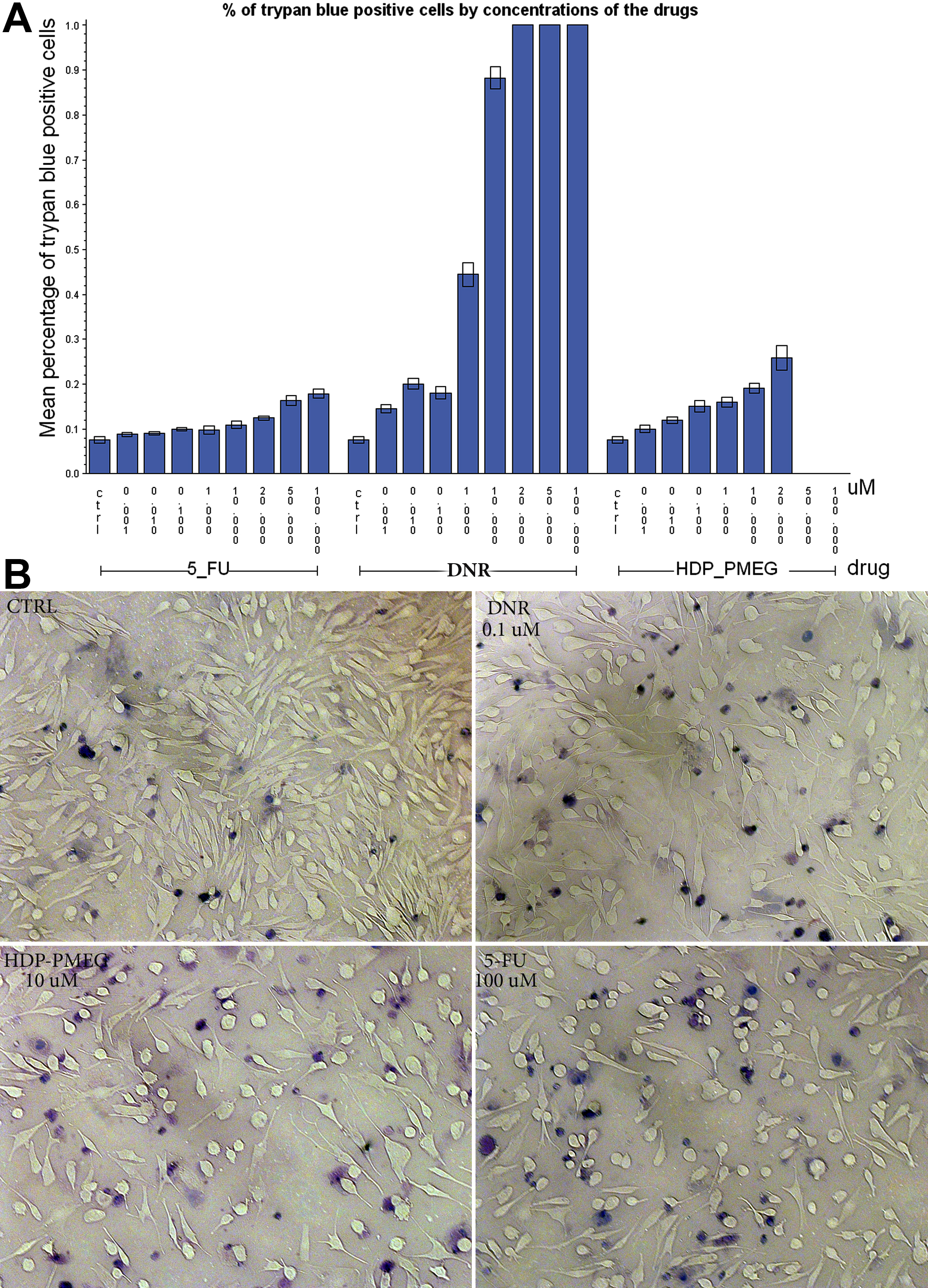
 Figure 4 of Hou, Mol Vis 2011; 17:627-637.