Figure 4. Immunohistochemical findings (MMP9 and MMP2). A-D: Consecutive sections from the case 1 sample. A, B: Intense MMP9 expression was seen in the stroma (A, B) and epithelium (B) near the perforation. C: MMP2 expression was not observed. E: Conjunctiva sample from a cGVHD patient showing intense MMP9 expression (positive control). F: Control conjunctiva sample (negative control).
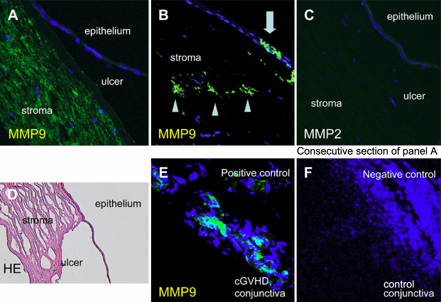
 Figure 4 of
Inagaki, Mol Vis 2011; 17:598-606.
Figure 4 of
Inagaki, Mol Vis 2011; 17:598-606.