Figure 3. Immunohistochemical findings (CD4, CD8, and CD68). A: CD68 expression at the margin of the corneal ulcer from case 4 sample. B, C: CD8+ (B) and CD4+ (C) T cells were not detected in this sample. E, F: Control samples for CD8 (E) and CD4 (F) immunostaining. D: Control sample. CD68 immunostaining.
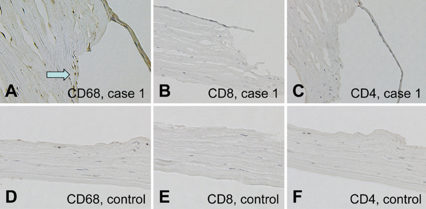
 Figure 3 of
Inagaki, Mol Vis 2011; 17:598-606.
Figure 3 of
Inagaki, Mol Vis 2011; 17:598-606.