Figure 2. Pathological findings. A, B: Case 4 frozen tissue section (cornea, hematoxylin & eosin staining, original magnification 40×, 100×). B: Magnified view of boxed area with an arrow in A. Corneal tissue near the perforation. C, D: Frozen tissue section (cornea, hematoxylin & eosin staining, original magnification 100×, 200×) Corneal tissue near the
perforation. The epithelium neighboring the corneal ulcer showed partial thinning (arrow).
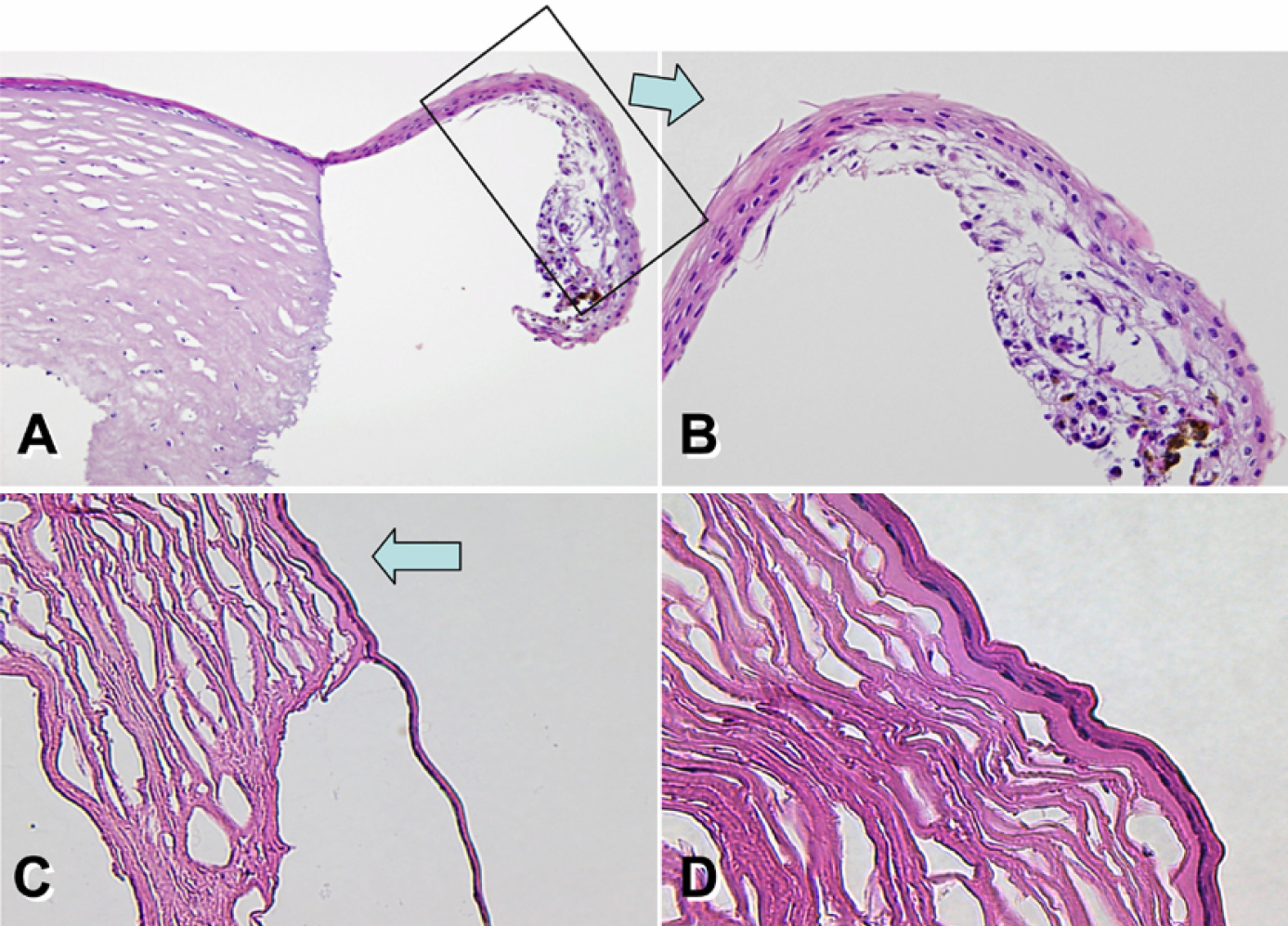
 Figure 2 of
Inagaki, Mol Vis 2011; 17:598-606.
Figure 2 of
Inagaki, Mol Vis 2011; 17:598-606.