Figure 1. Clinical findings of perforated cornea before and after treatment with deep anterior lamellar keratoplasty or glue. A, B: Case 1 photographs of the perforated cornea upon admission (left eye). A: The perforation was located in the paracentral cornea. The size of the perforation was 0.5 mm×0.5 mm. The surrounding ulcer
was 2.0 mm×1.5 mm. B: Preoperative photo showing a positive Seidel test. C: Photograph of the cornea after deep anterior lamellar keratoplasty. Five months later, the corneal graft remained stable
and transparent. D, E: Case 2 slit lamp photograph taken upon admission (right eye). D: The perforation was in the paracentral cornea and was 0.5 mm×1.0 mm. E: Photograph showing the paracentral corneal perforation plugged by the iris. F: Slit lamp photograph after the operation. One year after deep anterior lamellar keratoplasty, the corneal graft was stable
and transparent. G, H: Case 3 slit lamp photograph taken upon admission (left eye). G: The perforation was inferior to the center of the cornea and was 0.5 mm×0.5 mm. An ulcer (2.0 mm×1.0 mm) was observed inferiorly.
H: The anterior chamber was maintained by the prolapsed iris. I: Slit lamp photograph 3 months after healing of the perforated cornea without surgical intervention. J: Case 4 slit lamp photograph taken upon admission (right eye). The perforation was located inferior to the center of the
cornea. The cornea was 3.5 mm×1.5 mm, and the perforation was 2.0 mm×1.0 mm. The Seidel test was positive.
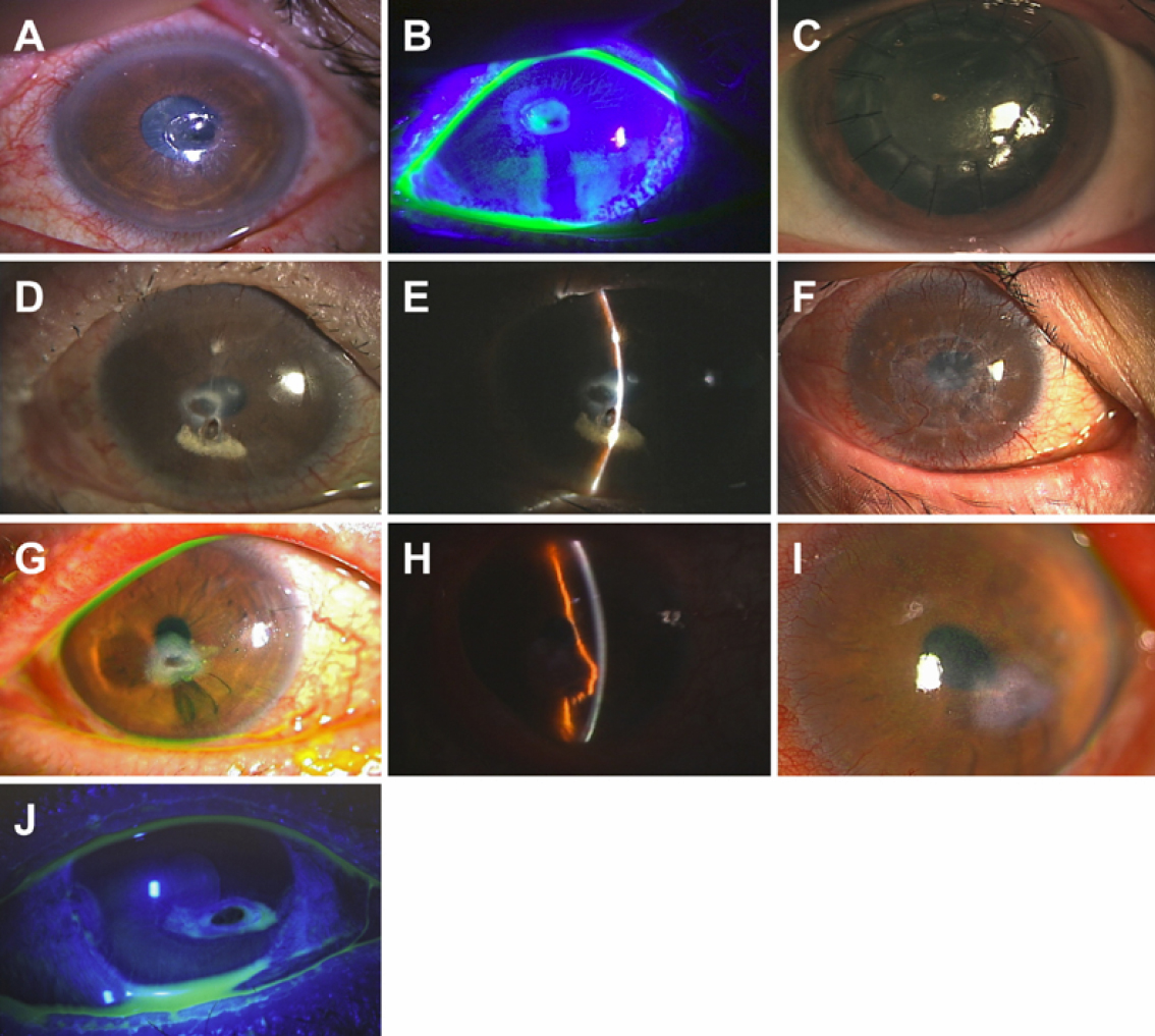
 Figure 1 of
Inagaki, Mol Vis 2011; 17:598-606.
Figure 1 of
Inagaki, Mol Vis 2011; 17:598-606.