Figure 3. Co-localization of F-actin and FRMD7. HEK293T cells transiently transfected with EGFP-tagged wild-type FRMD7 and FRMD7 mutants,
c.781C>G and c.886G>C, (shown in green) were stained with TRITC-conjugated rhodamine–phalloidin fluorescein (red). All three
fusion proteins exhibited a diffuse localization pattern in the cytoplasm, and also in actin-rich regions of the HEK293T cells
(arrows). However, HEK293T cells expressing the FRMD7 mutant, c.1003C>T, fused to EGFP, primarily exhibited localization of
the fusion protein to the nucleus and did not co-localize with F-actin (merge). Scale bars: 20 μm.
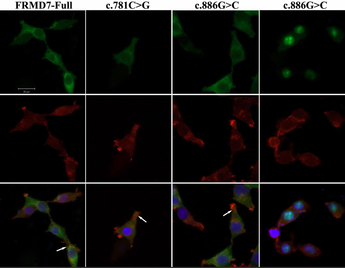
 Figure 3 of
Pu, Mol Vis 2011; 17:591-597.
Figure 3 of
Pu, Mol Vis 2011; 17:591-597.