Figure 2. Subcellular localization of full-length FRMD7 versus various domains of FRMD7. HEK293T cells transiently transfected with
EGFP-fusion proteins of FRMD7 (green) were stained with DAPI (blue) and imaged. Full-length FRMD7 was diffusely localized
to the cytoplasm, while the NH2-terminal FERM domain (aa 1–279), and the FERM domain plus the FA domain, primarily localized to the nucleus (arrows). Constructs
containing the COOH-terminal region of FRMD7 (e.g., △FERM and △FERM+FA) exhibited an expression pattern similar to that of
full-length FRMD7, however, the expression level was decreased and some aggregates of these truncated proteins were detected
in the cytoplasm. The results obtained from Neuro-2a cells (not shown) was the same as HEK293T cells. FERM: four-point-one,
ezrin, radixin, moesin; FERM+FA: FERM domain and FERM Adjacent domain; △FERM: truncated NH2-terminal FERM domain; △FERM+FA; truncated NH2-terminal FERM domain and FERM Adjacent domain. Scale bars: 20 μm.
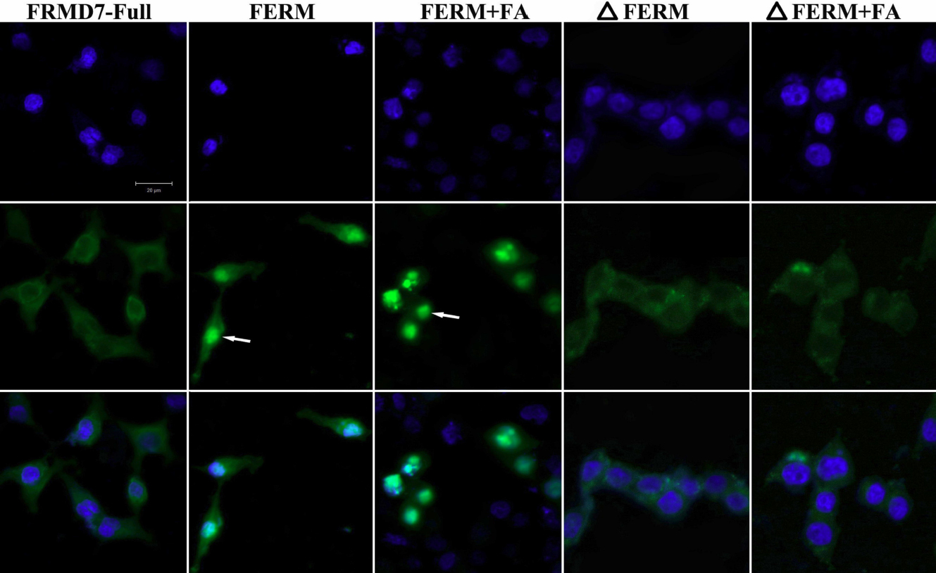
 Figure 2 of
Pu, Mol Vis 2011; 17:591-597.
Figure 2 of
Pu, Mol Vis 2011; 17:591-597.