Figure 5. Comparison of two-photon
microscopy (2PM) and conventional histology. A: A true radial
histological section. B: A virtual radial-section calculated
using data from the multiple z-sections imaged. The angle of this
radial section is tilted 60° with respect to the face of the sclera,
giving a diagonal cross section of the eye. The iris root (IR),
Schlemm’s canal (SC, white arrows), collector channel (red lines and
*), and TM (dotted lines) are visible in both images. The end of SC
closest to the IR is visible in B, the 2PM image, but partially
collapsed against the TM in A, the histological section. The
connection between the collector channel and SC, outlined by red in the
2PM image in B, is also not visible in the histological section
in A. Scale bar=100 µm.
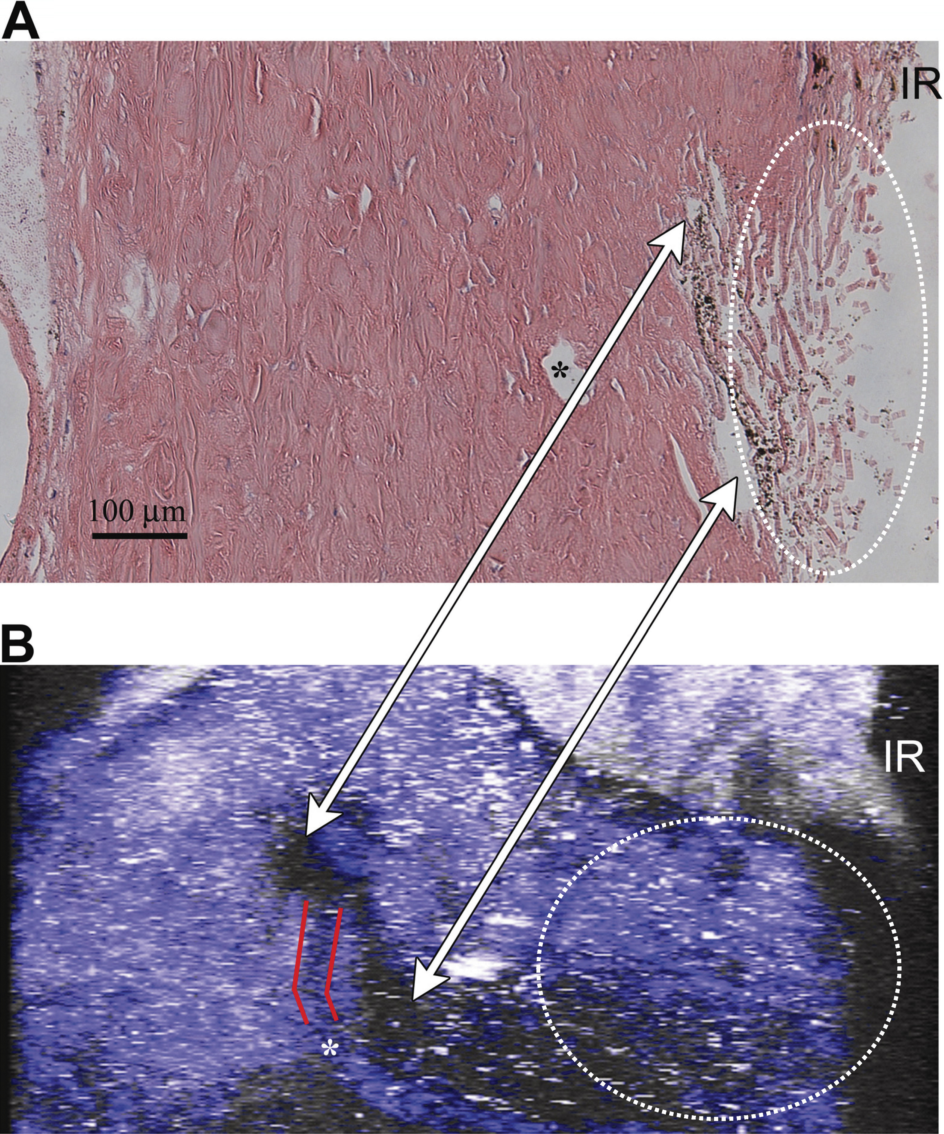
 Figure 5 of Ammar, Mol Vis 2011; 17:583-590.