Figure 4. Serial sections of the TM region
imaged by 2 photon microscopy. Sections shown here are indicated by the
distance from the edge of the tissue block (in microns). The Schlemm’s
Canal (SC) opening is apparent in many of the sections. Also prevalent
is a ~50 µm round structure, representing a collector channel (CC),
visible from the 900 µm section through the 2400 µm section. Black
bar=100 µm.
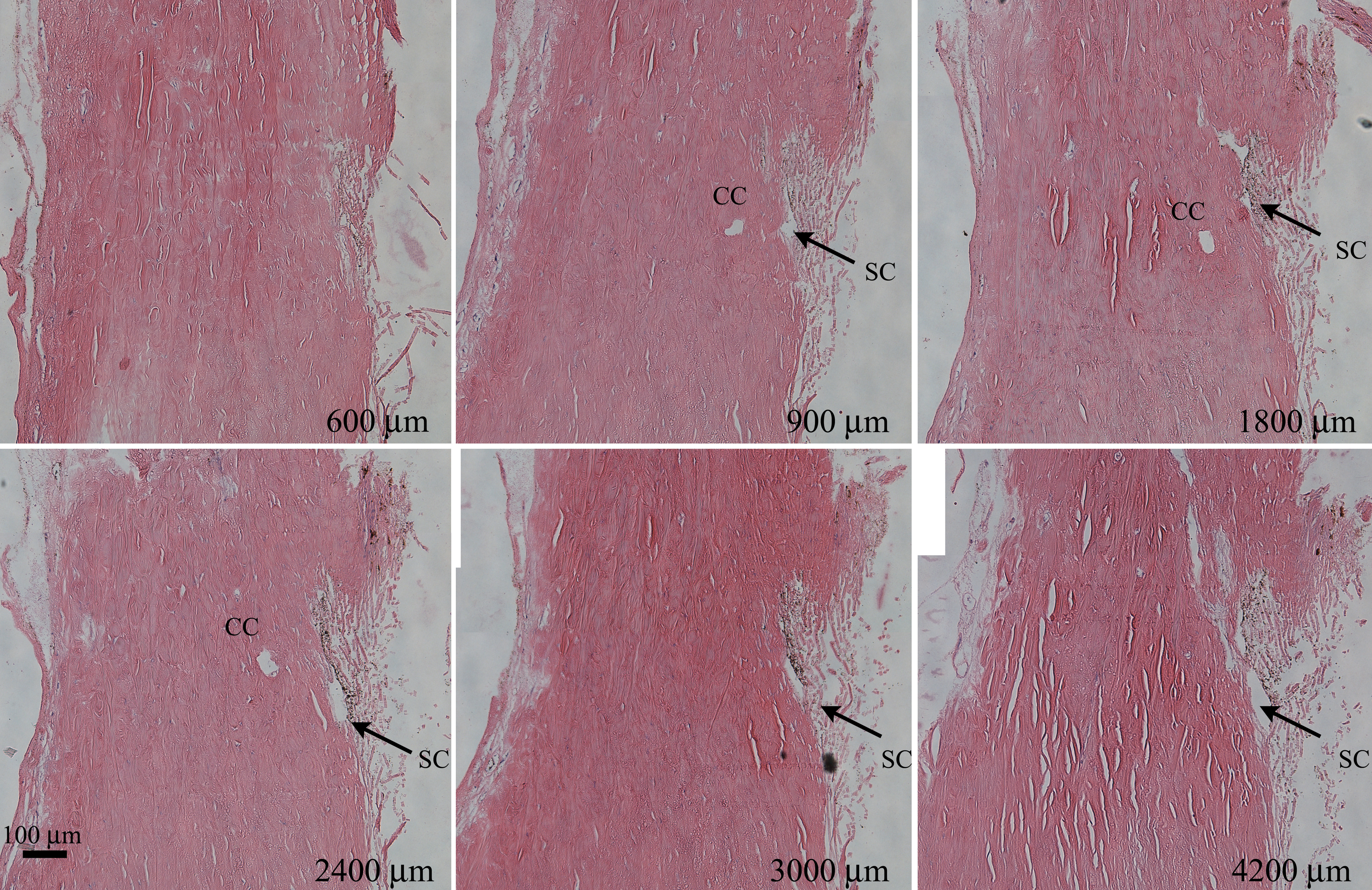
 Figure 4 of Ammar, Mol Vis 2011; 17:583-590.