Figure 1. A histological section of the
same human trabecular meshwork (TM) region imaged by 2PM. The paraffin
section was stained with hematoxylin and eosin and placed alongside a
schematic outline. The TM region is clearly visible in the lower
right-hand edge of each section (toward the anterior chamber). The TM
region shows darkly-stained endothelial cells populating a pink-stained
fibrous tissue region of the eye. Schlemm’s canal is visible proximal
to the TM region (outlined in red in schematic). The scleral spur and
iris remnant are also labeled in the schematic, as are multiple opens
spaces formed during processing the tissue for histology (artifacts).
Black bar=100 µm.
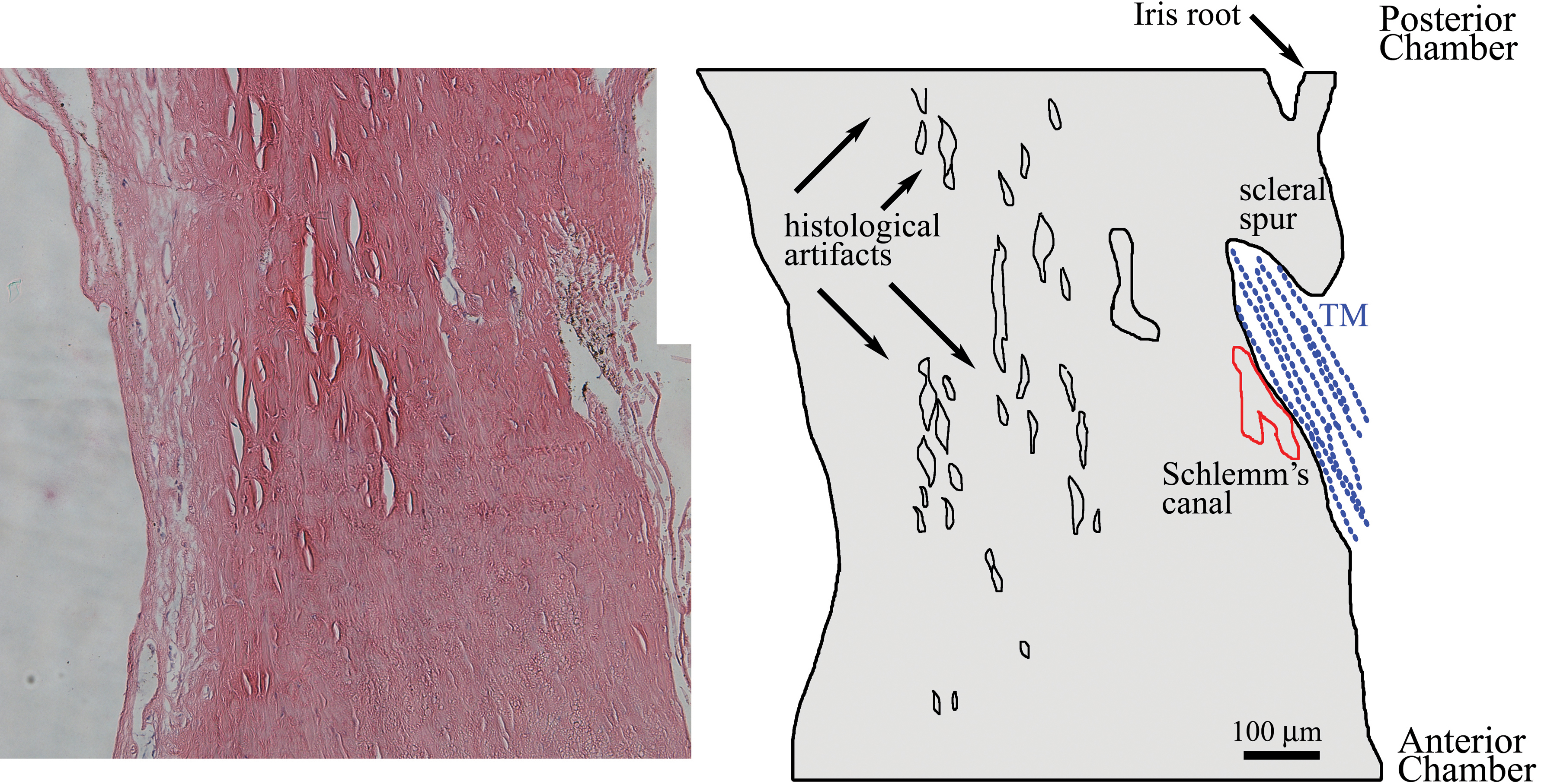
 Figure 1 of Ammar, Mol Vis 2011; 17:583-590.