Figure 2. Angiogenin localization in human
eyes. Labeling of angiogenin is shown in a normal macular choroid (A),
in
which angiogenin is expressed on the endothelial cells of the
choriocapillaris and large vessels, as well as localized to
intercapillary pillars. Modest anti-angiogenin labeling is detected in
some extramacular drusen (C and D, asterisks). In some
donors, approximately 50% of the hard drusen showed weak
immunoreactivity (C). Minor labeling of a hyaline druse (D,
asterisk)
was observed in an 83-year-old female with neovascular age
related macular degeneration (AMD), compared to an adjacent section in
which the primary antibody was omitted (E). Endothelial cells in
a neovascular membrane (F) from the macula of human donor eye
with advanced AMD were also reactive with anti-angiogenin antibody. The
presence of angiogenin in the neovascular membrane vessels indicates
that it may play a role in the pathophysiology of these cells in AMD.
Controls in which primary antibodies are omitted are shown for normal
tissue (B), drusen (E), and neovascular tissue (G).
Scale
bar represents 50 μm.
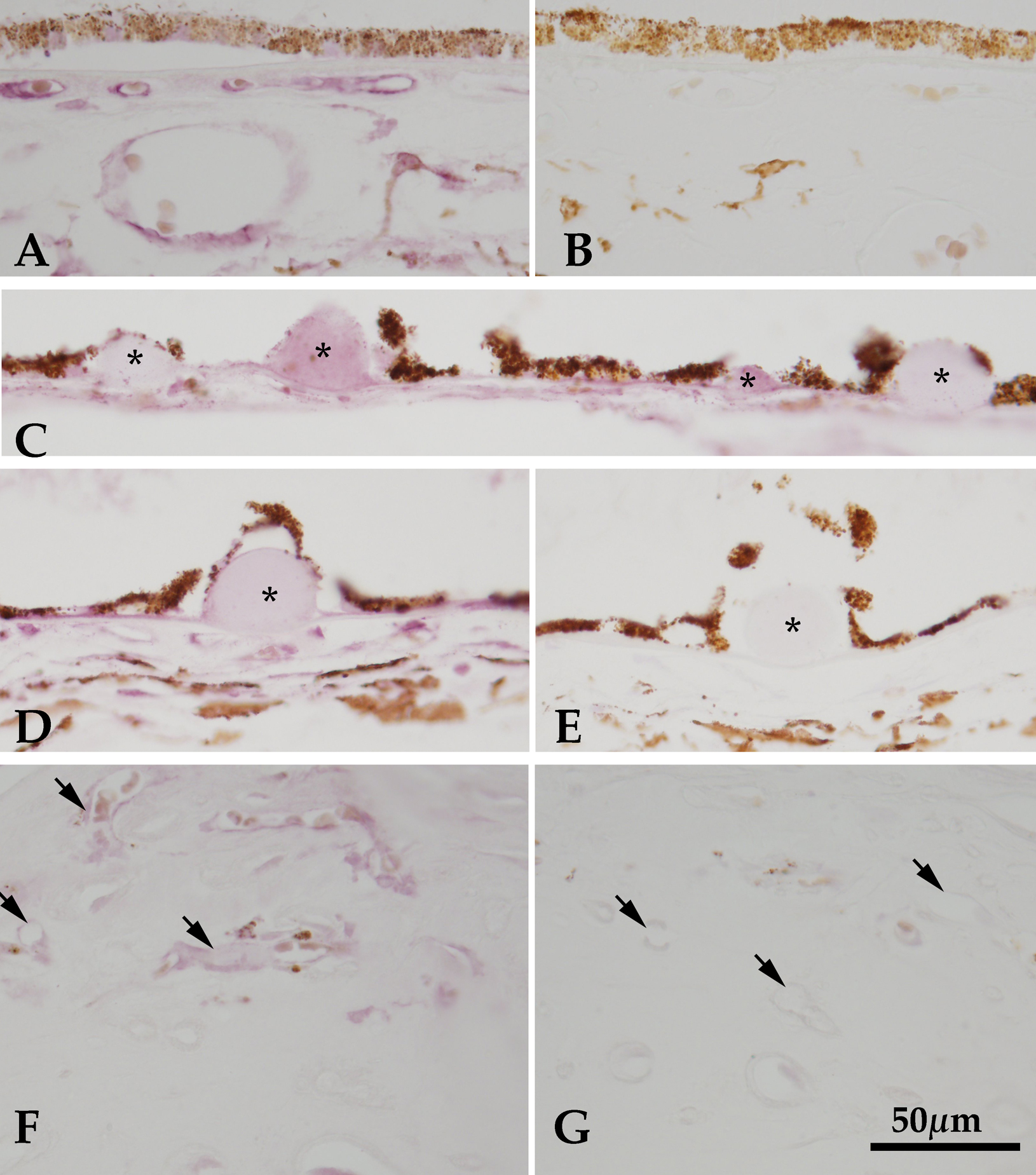
 Figure 2 of Skeie, Mol Vis 2011; 17:576-582.