Figure 7. Differentiation of human
pluripotent stem cells toward retinal pigment epithelium (RPE) cells
under defined culture conditions, RPEregES. All represented images are
from human embryonic stem cell (hESC)-RPE Regea 08/023. A:
Reverse transcription (RT)–PCR analysis of typical genes for retinal/
RPE development expressed by undifferentiated hESC (Regea 08/023),
human foreskin fibroblast (hFF) feeder cells, and putative hESC-RPE on
D7 and D44. Expression of B: Microphthalmia-associated
transcription factor (MITF), B: Cellular retinaldehyde-binding
protein (CRALBP), and E,G: RPE65 on D83. F: For
cell morphology, F-actins were stained using phalloidin. H:
Proliferative activity was studied by Ki67 staining together with tight
junction protein anti-zonula occludens (ZO)-1 in hESC-RPE. I:
Vertical confocal sections showing apical localization of Na+/K+ATPase
(green)
and basolateral localization of Bestrophin (red). Nuclei
stained with 4',6-diamidino-2-phenylindole (DAPI). Images B-D
were taken with an Olympus BX60 microscope (Olympus, Tokyo, Japan)
using a 60× oil immersion objective, scale bar 20 μm. Images E-I
were taken with an LSM 700 confocal microscope (Carl Zeiss) using a 63×
oil immersion objective, scale bar 20 μm.
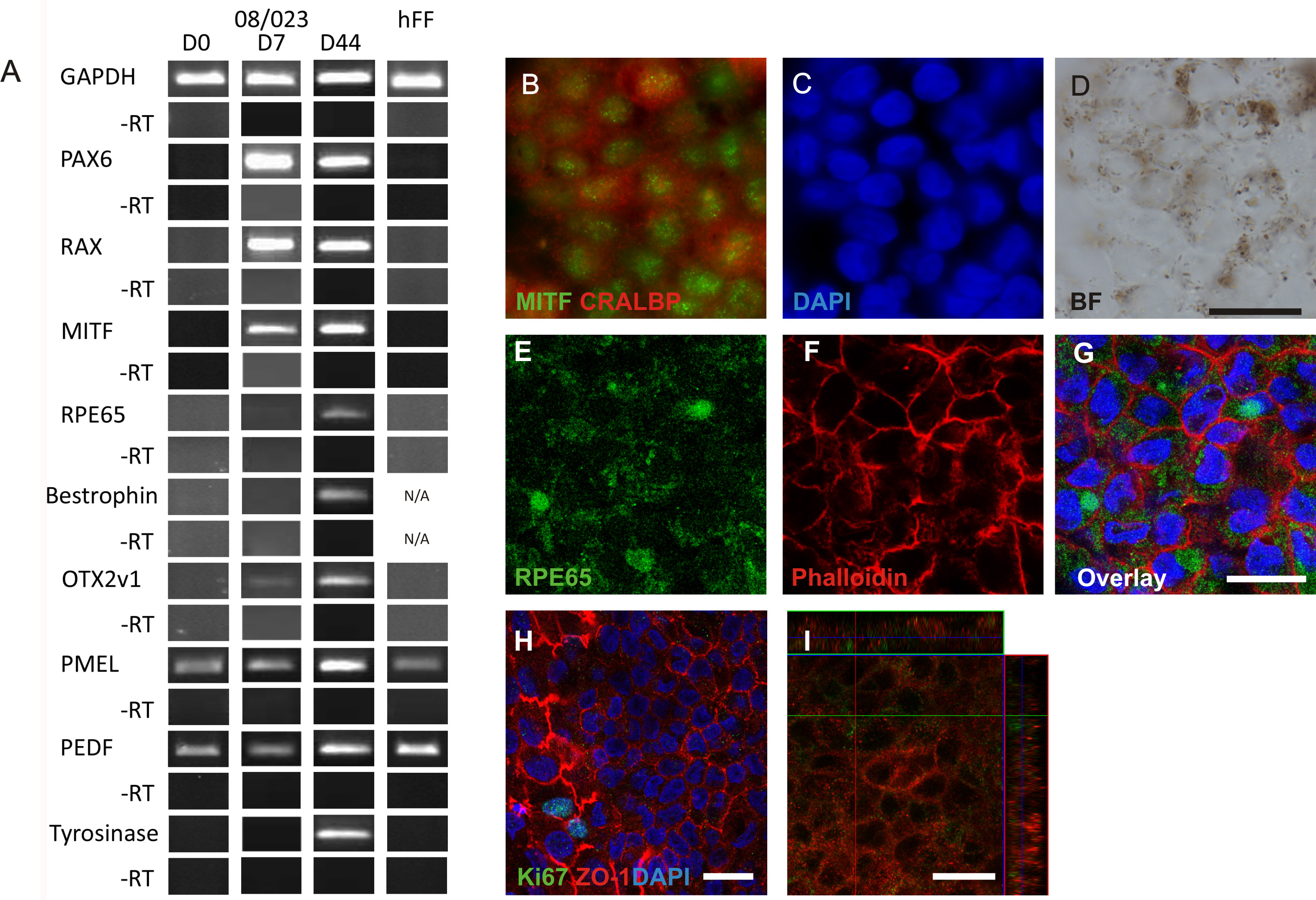
 Figure 7 of Vaajasaari, Mol Vis 2011; 17:558-575.