Figure 5. Morphology and gene expression
analysis of manually selected and long-term cultured human embryonic
stem cell (hESC)-retinal pigment epithelium (RPE; Regea 08/023) and
hiPSC-RPE (FiPS 5–7) cells. A: Bright-field micrograph of
hESC-retinal pigment epithelium (RPE) and human induced pluripotent
stem cell (hiPSC)-RPE cells cultured for 136 days on Collagen IV. The
cells have acquired a cobblestone morphology and a high degree of
pigmentation, which is typical of RPE cells. Low magnification images
were captured with a Nikon Eclipse TE2000-S phase contrast microscope
(Nikon Instruments Europe B.V. Amstelveen, The Netherlands) and higher
magnification images with an Olympus BX60 microscope (Olympus, Tokyo,
Japan) using a 60× oil immersion objective. Scale bar 20 μm. B:
Reverse transcription (RT)–PCR analysis showing the expression of optic
vesicle, optic cup, RPE, neural crest melanocyte, pluripotent stem
cell, mesoderm and endoderm marker genes by undifferentiated cells
(D0), human foreskin fibroblast (hFF) feeder cells, and putative RPE
cells (D196) from Regea 08/023 and FiPS 5–7 cells. N/A=not analyzed. C:
Relative
OCT3/4 expression between undifferentiated FiPS 5–7 and
putative hiPSC-RPE after 196 days of differentiation.
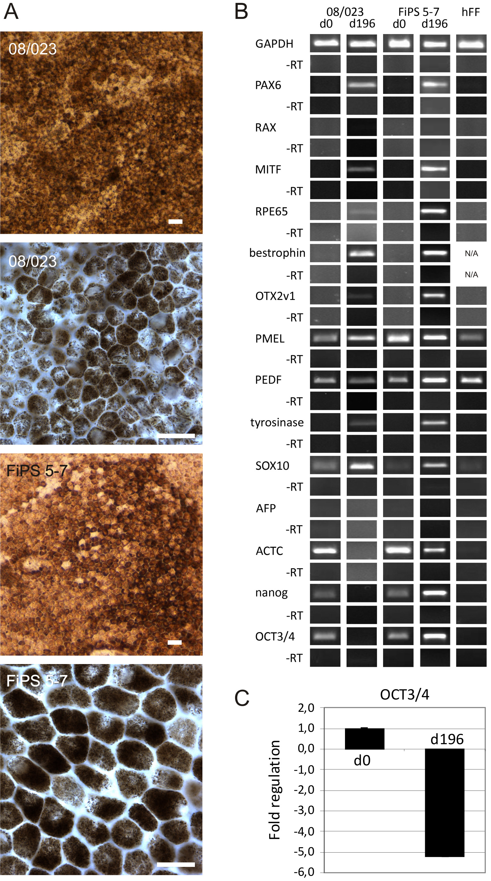
 Figure 5 of Vaajasaari, Mol Vis 2011; 17:558-575.