Figure 3. Immunofluorescence staining of
human embryonic stem cell (hESC; Regea 08/023)- and human induced
pluripotent stem cell (hiPSC; FiPS 5–7)-derived retinal pigment
epithelium cells revealing maturation stage after 83 days of
differentiation. Cellular retinaldehyde-binding protein (CRALBP) and
microphthalmia-associated transcription factor (MITF) localization in A-C:
manually
selected hESC-RPE cells and D-F: hiPSC-retinal pigment
epithelium (RPE) cells. G, I: RPE65 expression in
hESC-RPE and J, L: hiPSC-RPE. H, K: For
cell morphology, F-actins were stained using phalloidin. Tight junction
protein anti-zonula occludens (ZO)-1 and proliferation marker Ki67
localization in M: hESC-RPE cells and N: hiPSC-RPE
cells. Nuclei were stained with 4',6-diamidino-2-phenylindole (DAPI).
Images A-F were taken with an Olympus BX60 microscope
(Olympus, Tokyo, Japan) using a 60× oil immersion objective, scale bar
20 μm. Images G-N were taken with an LSM 700 confocal
microscope (Carl Zeiss) using a 63× oil immersion objective, scale bar
20 μm.
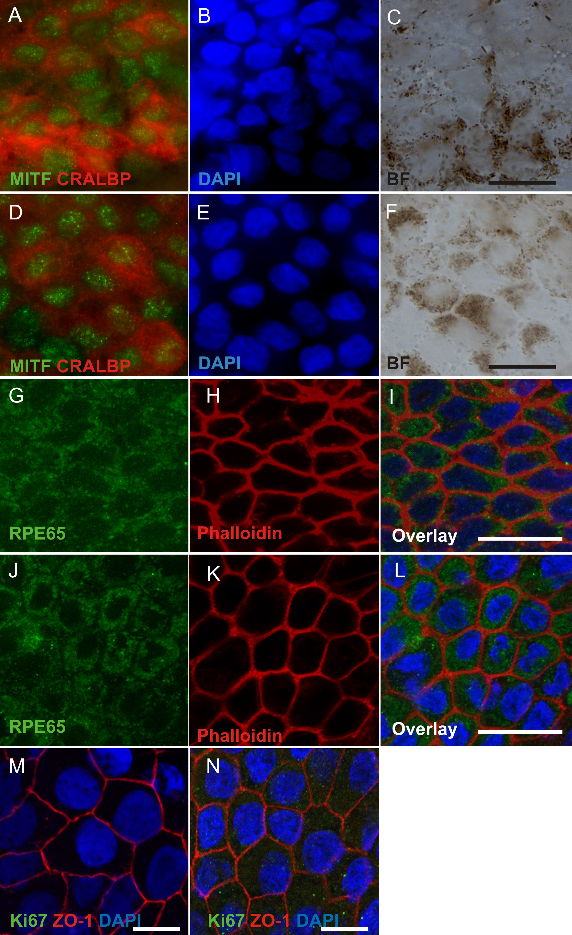
 Figure 3 of Vaajasaari, Mol Vis 2011; 17:558-575.