Figure 3. Ophthalmological findings in patients from the two families. A: Photograph of anterior segment of patient III-4 of 85 with complete absence of iris and the progressing dense congenital
cataract. B: Fundus images of patient III-4 showed late-stage glaucomatous cupping of the optic disc. C: Complete hypoplasia of the iris and congenital cataract were observed in patient IV-7 of family 85. D: Fundus images of patient IV-7 showing foveal hypoplasia. E: Photograph of anterior segment of patient II-4 of family 86 with complete absence of iris and congenital cataract. F: Fundus image of patient II-4 showing a tessellated appearance. G: Photograph of the anterior segment of patient III-3 of family 86 with complete absence of iris and mild cataract. H: Fundus image of patient III-3 showing a normal foveal reflex.
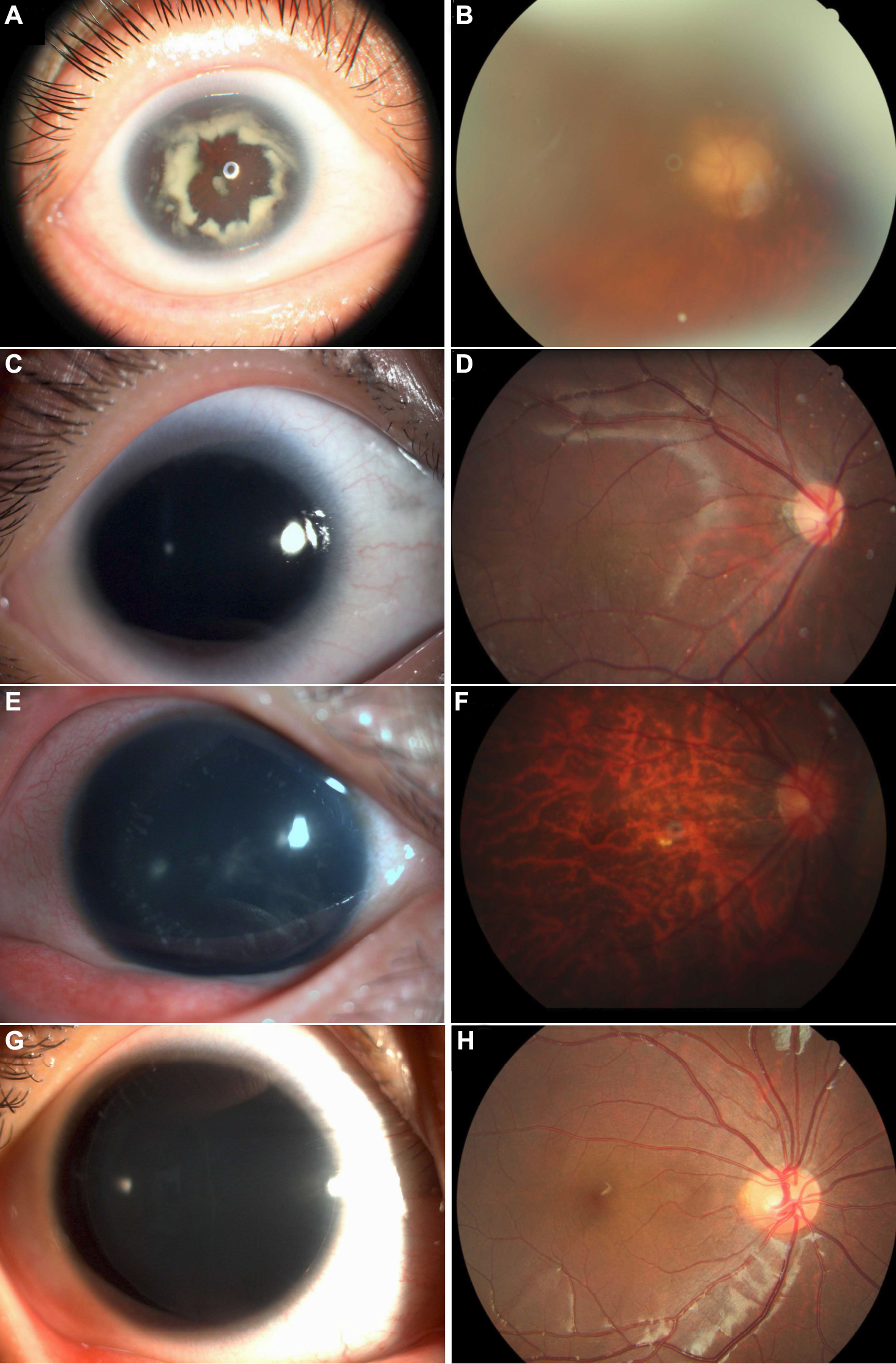
 Figure 3 of
Zhang, Mol Vis 2011; 17:548-557.
Figure 3 of
Zhang, Mol Vis 2011; 17:548-557.