Figure 4. Effects of EGCG on IL-1β-induced
phosphorylated p38 and JNK levels in HCEpiC. Cells were cultured in
complete (HCGS-containing) medium, followed by basal medium for 18 h.
Cells were pre-treated with EGCG for 2 h. Cells were then treated with
IL-1β + EGCG for 30 min. Phosphorylated p38 and JNK levels were
determined by cell-based ELISA (upper panels) or western blotting
(lower panels). A: effect of EGCG on IL-1β-induced
phosphorylated p38 determined by cell-based ELISA; B: effect of
EGCG on IL-1β-induced phosphorylated JNK determined by cell-based
ELISA; C: effect of EGCG on IL-1β-induced phosphorylated p38
determined by western blotting, Upper panel: upper blot shows
phosphorylated p38. Lower blot is after stripping and probing with
total p38 antibody. Lower panel: densitometric analysis of
phosphorylated p38 normalized by total p38. D. effect of EGCG on
IL-1β-induced phosphorylated JNK determined by western blotting, Upper
panel: upper blot shows phosphorylated JNK. Lower blot is after
stripping and probing with total JNK antibody. Lower left panel:
densitometric analysis of phosphorylated p46 JNK normalized by total
p46 JNK; lower right panel: densitometric analysis of phosphorylated
p54 JNK normalized by total p54 JNK. For A and B, n=6,
For C and D, n=3–4. Representative blots are shown.
*versus IL-1β; p<0.05.
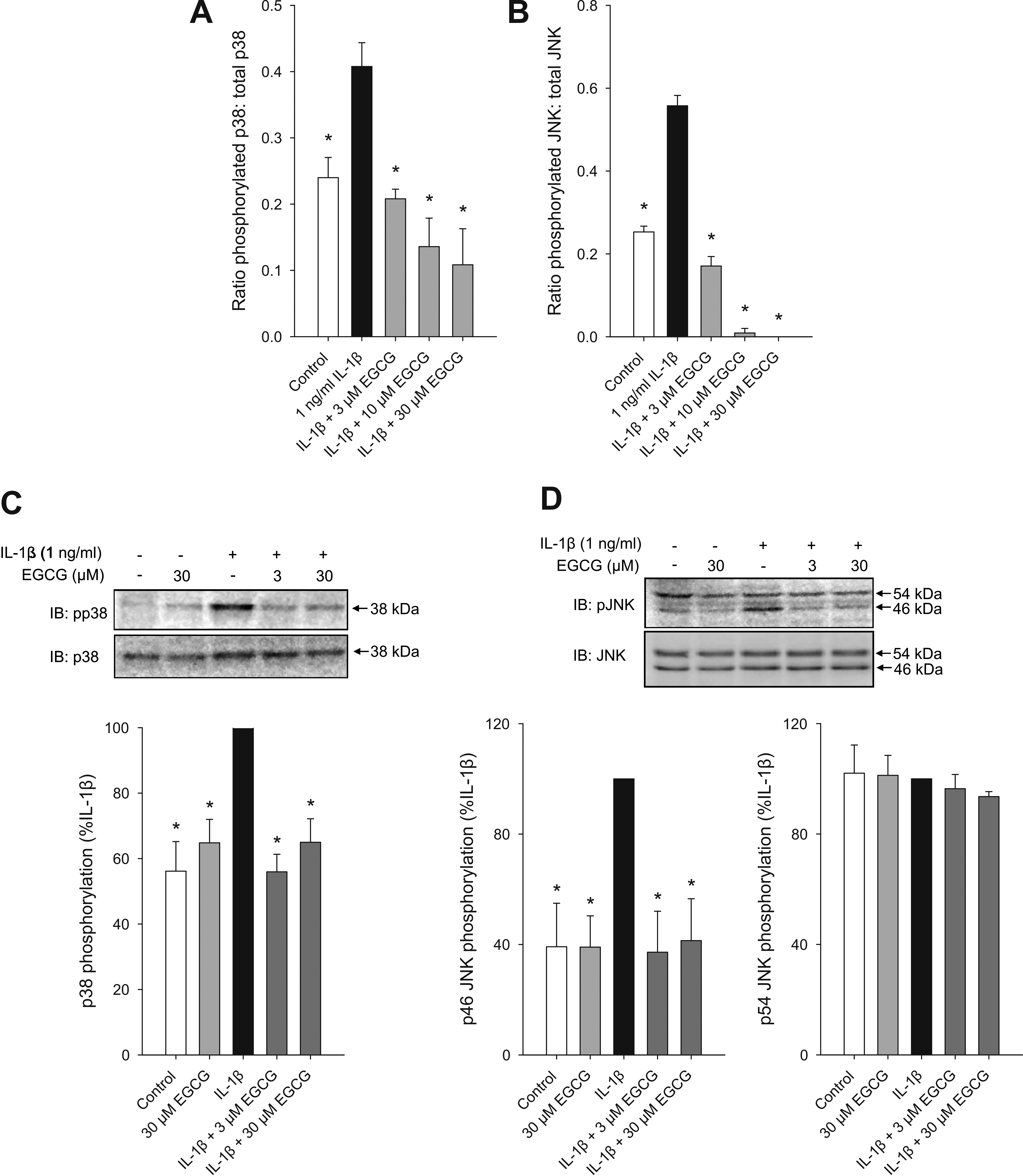
 Figure 4 of Cavet, Mol Vis 2011; 17:533-542.