Figure 6. Both vascular endothelial growth
factor-B167 and 186 increased retinal neovascularization in oxygen
induced ischemic retinopathy. At postnatal day 7 (P7), pups received an
intravitreal injection of AAV-VEGF-B167, AAV-VEGF-B186,
or AAV-GFP vectors, then were placed in a 75% oxygen box for 5
days and returned to room air at P12 for another 5 days. At P17, mice
were sacrificed, retinal-whole mounts prepared, and retinal
neovascularization quantified as described in Methods. Images from A
to C are representative retina whole mounts from mice that
received AAV-GFP (A), AAV-VEGF-B167 (B),
or AAV-VEGF-B186 (C). Original magnification was ×5. All
images were taken under the same conditions for optimal comparison. D:
Statistical analysis indicated that both isoforms of VEGF-B
significantly increased the retinal neovascularization area compared
with the GFP control group. The graph shows that AAV-VEGF-B167
and 186 promote increased retinal neovascularization in the
oxygen induced ischemic retinopathy model. Data are expressed as
mean±SEM.
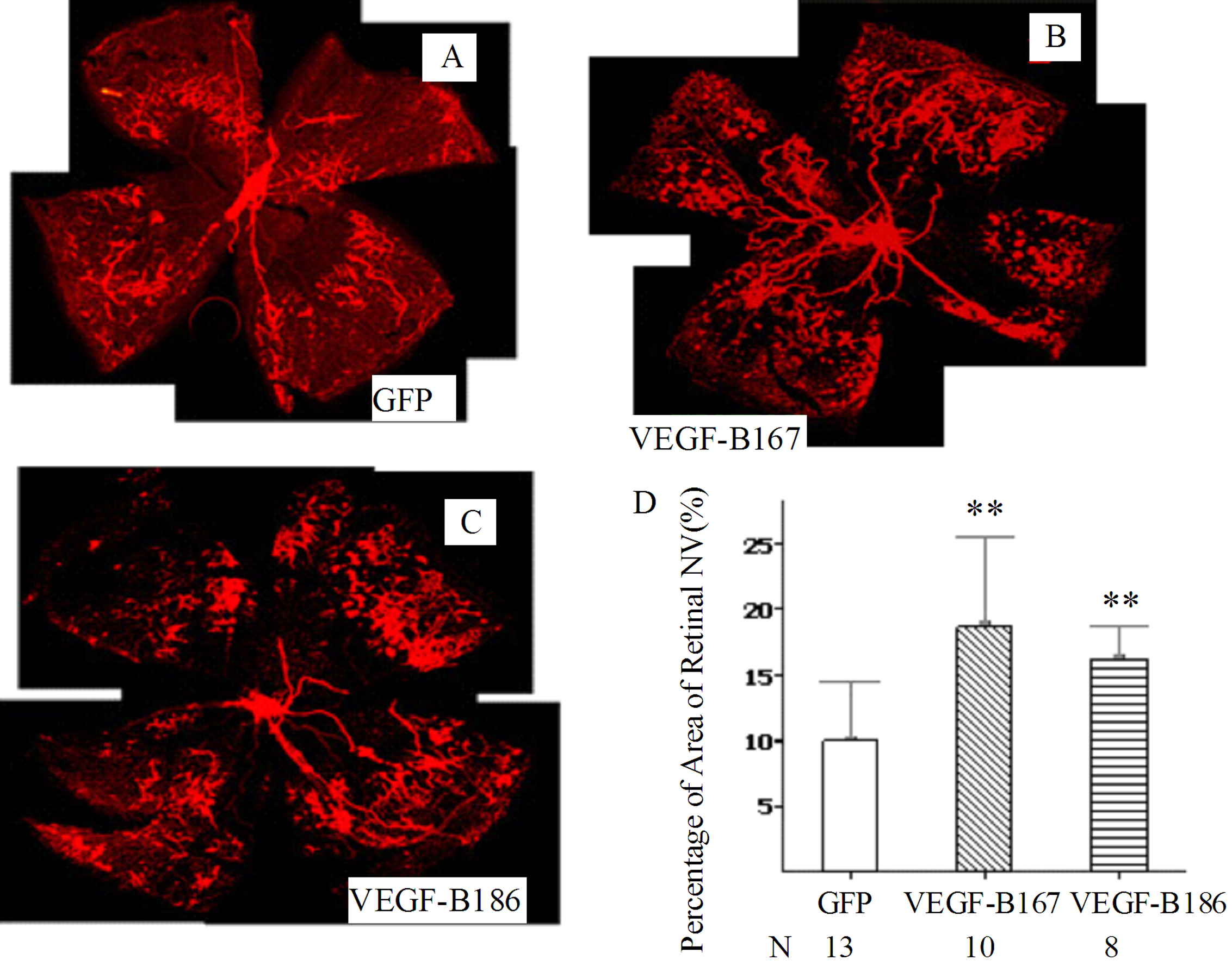
 Figure 6 of Zhong, Mol Vis 2011; 17:492-507.