Figure 5. Representative images show
transgene expression after intravitreous delivery of recombinant
adeno-associated virus-green fluorescent protein (AAV-GFP) in
the
oxygen induced ischemic retinopathy (OIR) model. At postnatal day 7,
pups received an intravitreal injection of AAV-GFP vector and
were then placed in a 75% oxygen box for 5 days. Thereafter, pups were
returned to room air for another 5 days. At day 10 after injection
(postnatal day 17), pups were killed. Choroidal flatmounts (A, B)
and
cryosections
(C, D) were prepared. Green fluorescent
protein expression was directly observed using fluorescence microscopy.
Original magnifications were as follows: A: ×5, B: ×40 C,
D: ×20 .
The figure shows increased expression of the reporter gene, GFP.
The
following
layers are illustrated: RPE: retinal pigmental
epithelium, OS: outer segment, OPL: outer plexiform layer, INL: inner
nuclear layer, IPL: inner plexiform layer, GCL: ganglion cell layer.
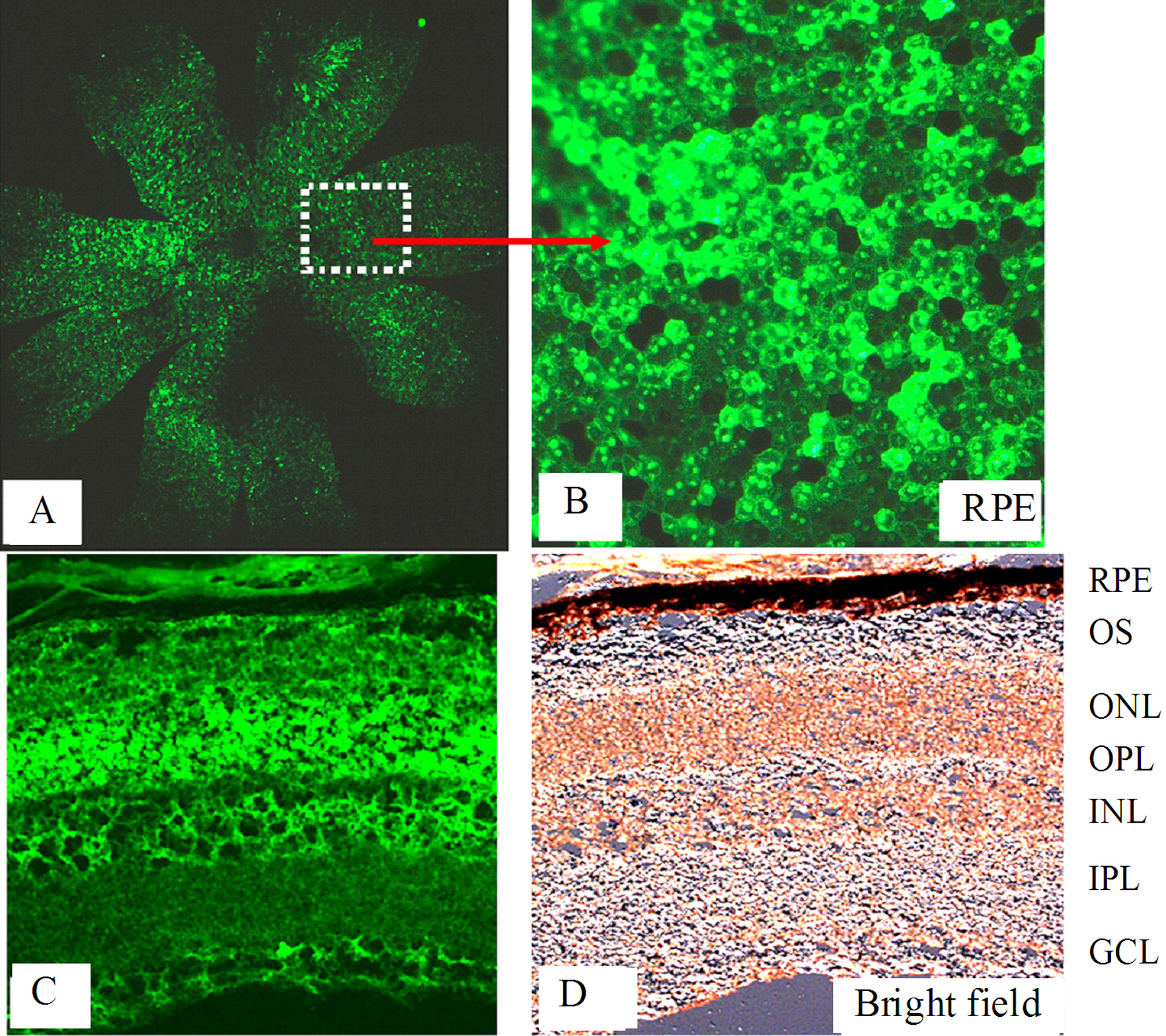
 Figure 5 of Zhong, Mol Vis 2011; 17:492-507.