Figure 4. Increased choroidal
neovascularization caused by overexpression of adeno-associated
virus-vascular endothelial growth factor (AAV-VEGF)-B167
and 186 at rupture sites in Bruch’s membrane. A–C:
Two
weeks
after subretinal injection of AAV-VEGF-B167(A),
AAV-VEGF-B186(B), or AAV-GFP(C), mice
underwent laser photocoagulation that ruptured Bruch’s membrane in
three locations in each eye. Fourteen days later, mice were perfused
with fluorescein-labeled dextran, and choroidal flatmounts were
prepared and examined by fluorescence microscopy. Compared to eyes
treated with a subretinal injection of AAV-GFP(C), the
area of choroidal neovascularization at Bruch’s membrane rupture sites
was significantly increased in eyes given a subretinal injection of AAV-VEGF-B167
(A, D) or 186 (B, D). The arrows
define the limits of the choroidal neovascularization. D shows
that the area of choroidal neovascularization (CNV), expressed in mm2,
is
significantly
increased following a subretinal injection of AAV-VEGFB-B167
or 186. Data are expressed as mean±SEM. Statistical comparisons
were
made by unpaired t test (*p<0.05). Images A to C
have the same scale. Scale bar represents 100 μm.
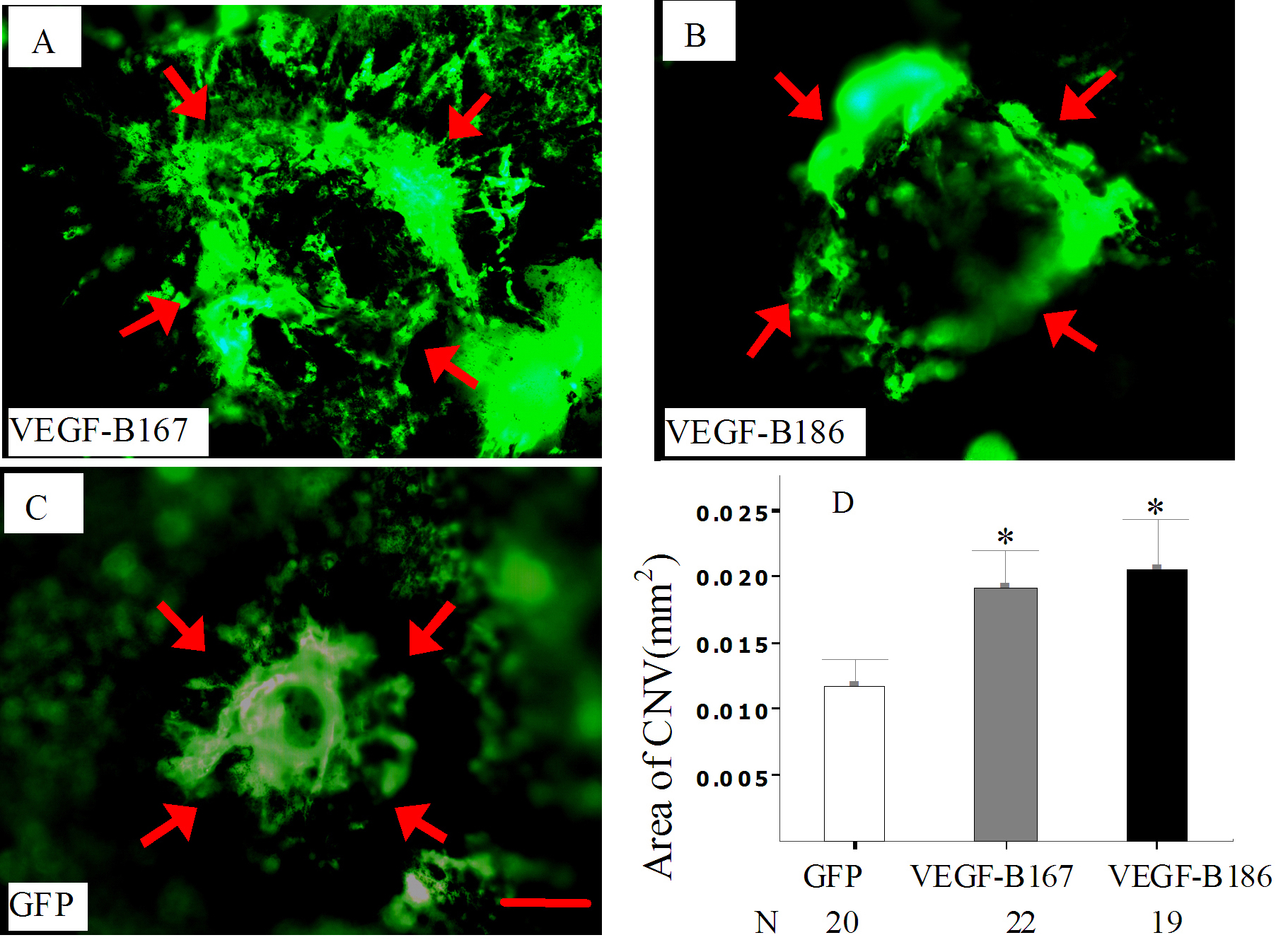
 Figure 4 of Zhong, Mol Vis 2011; 17:492-507.