Figure 2. No obvious induction by vascular
endothelial growth factor (VEGF)-B167 or 186 of inflammatory response 4
weeks after subretinal injection of adeno-associated virus (AAV)-VEGF-B167
and 186 in C57BL/6J mice. A–C: Hematoxylin and
eosin staining; D–G: Leukostasis assay indicated that
neither AAV-VEGF-B167 (E, G) nor AAV-VEGF-B186
(F, G) caused significant retinal leukostasis compared
to the AAV-GFP control (D). Data are presented as
mean±SEM.
Sample sizes for GFP and VEGF-B167 are 10 and for VEGF-B186 is 8. For
GFP versus VEGF-B167, p=0.13; for GFP versus VEGF-B186, p=0.3. H:
An
image
for comparison showing vascular endothelial growth factor
(VEGF)-A-induced leukostasis 6 h after intravitreal administration of
1×10⁻⁶ M VEGF-A. Arrows are pointing to leukocytes adhering
to the vessel wall. Original magnification was ×20.
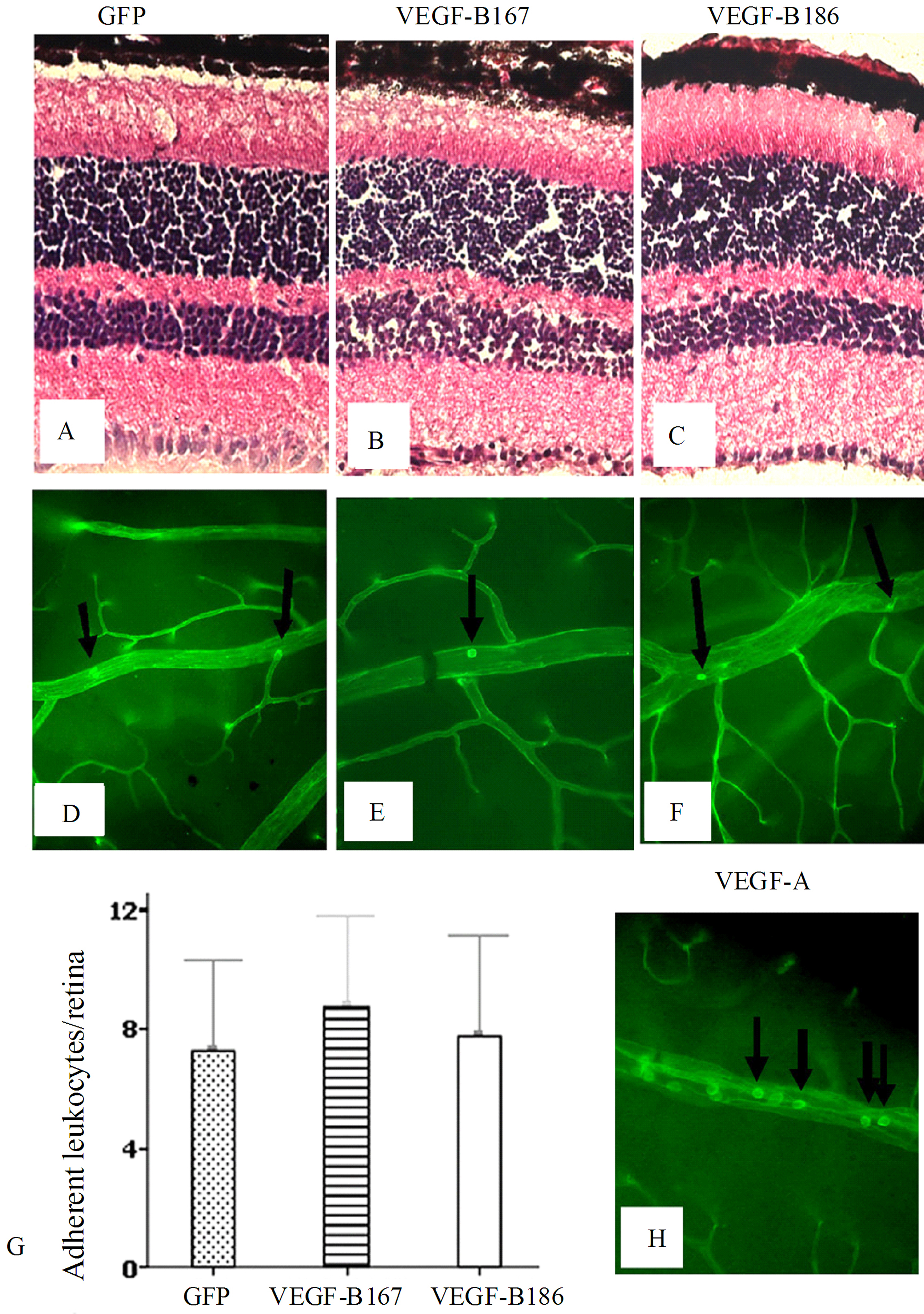
 Figure 2 of Zhong, Mol Vis 2011; 17:492-507.