Figure 1. This figure shows the transgene
expression of recombinant adeno-associated virus (rAAV) vectors in the
retinas of 4-week-old C57BL/6J mice, 4 weeks after subretinal injection
of approximately 1×10⁹ vgc recombinant adeno-associated
virus vectors. A: This shows the schematic representation of
the three rAAV vectors used in this study for transducing vascular
endothelial growth factor (VEGF)-B167, VEGF-B186,
and the marker GFP gene. Terminal repeats (TR), AAV terminal
repeats; cytomegalovirus (CMV), human cytomegalovirus immediate–early
promoter; pA, polyadenylation site. B–D: Expression of
GFP protein is demonstrated in flatmounts of the choroid (B),
retina (C), and a section of eye (D); note the greatest
expression of green fluorescent protein (GFP) in the retinal pigment
epithelium (RPE; B and D, red arrow) and the outer
retina (C and D, black arrow). E–G:
Immunofluorescence staining indicated that the VEGF-B167 (E) and
VEGF-B186 proteins (F) are more strongly expressed in retinas
from experimental groups—mainly in the RPE—than in retinas from the GFP
control group (G). Cell nuclei were stained with 4´,
6-diamidino-2´-phenylindole dihydrochloride. Images E–G
were taken under the same conditions for optimal comparison. H–I:
Real-time
PCR analysis of VEGF-B167 (H) and 186 (I)
expression shows that mRNA expression levels of VEGF-B167 and 186 in
the AAV-VEGF-B167 and 186 groups are significantly
higher than those in the AAV-GFP control group (in arbitrary
units normalized against mouse β-actin; *p<0.05, **p<0.001). Data
are espressed ±SEM and sample sizes were 3. B–G: Scale
bar represents 50 µm.
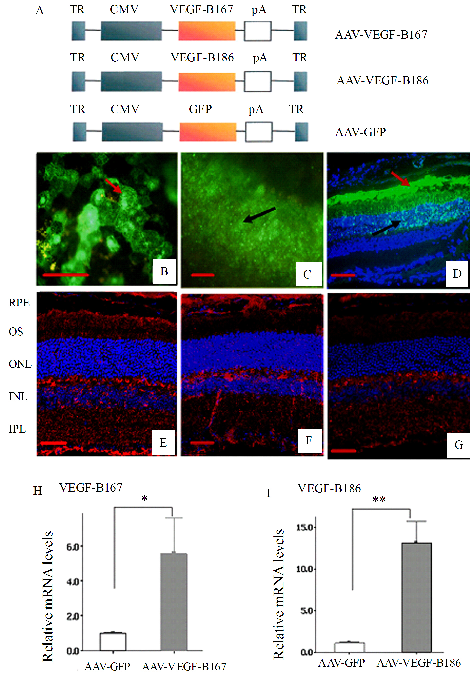
 Figure 1 of Zhong, Mol Vis 2011; 17:492-507.