Figure 1. In vitro culture of HCE cells. A:
HCE
cells 36 h after plating, showing the non-spread polygonal cell
morphology, i.e., corneal endothelial-like morphology. B: The
monolayer formed by HCE cells 6 weeks after primary culture initiation,
showing the plump polygonal cell morphology. C: Passage 101 HCE
cells, showing the co-existence of endothelioid and fibroblast-like
morphology. D: Passage 224 HCE cells, showing the co-existence
of elongated polygonal and fibroblast-like morphology. Scale bar: 50 μm.
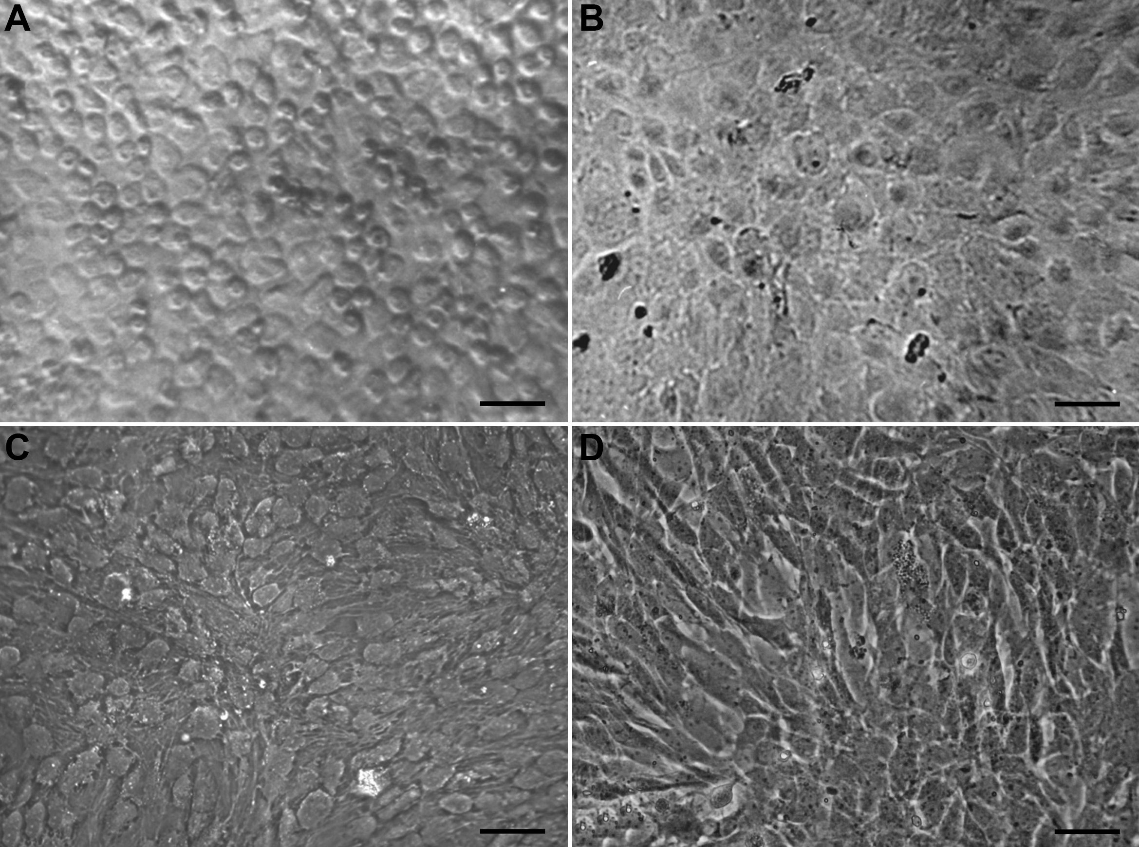
 Figure 1 of Fan, Mol Vis 2011; 17:469-480.