Figure 2. Caveolin 1 and 2 in the human
iridocorneal angle. Immunohistochemical detection of Caveolin 1 (A)
and
Caveolin 2 (B) in the iridocorneal angle. Caveolin 1
reactivity was mainly associated with the trabecular meshwork cells
lining the trabecular meshwork beams (Arrowhead) and Schlemm’s canal.
Caveolin 2 is mainly detected in Schlemm’s canal and the longitudinal
ciliary muscle. (SC: Schlemm’s canal, TM: Trabecular meshwork,
Magnification: (A) 400×, (B) 200×).
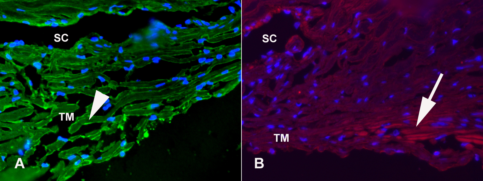
 Figure 2 of Kuehn, Mol Vis 2011; 17:430-435.