Figure 1. Caveolin 1 and 2 in the human
retina. Immunohistochemical detection of Caveolin 1 (A and B)
and
Caveolin 2 (C and D) in the human retina. Negative
controls with no primary antibody (B and D) revealed
only minor cross reactivity of the secondary antibodies. GCL=ganglion
cell layer, INL=Inner nuclear layer, ONL=outer nuclear layer, OS=Outer
segments.
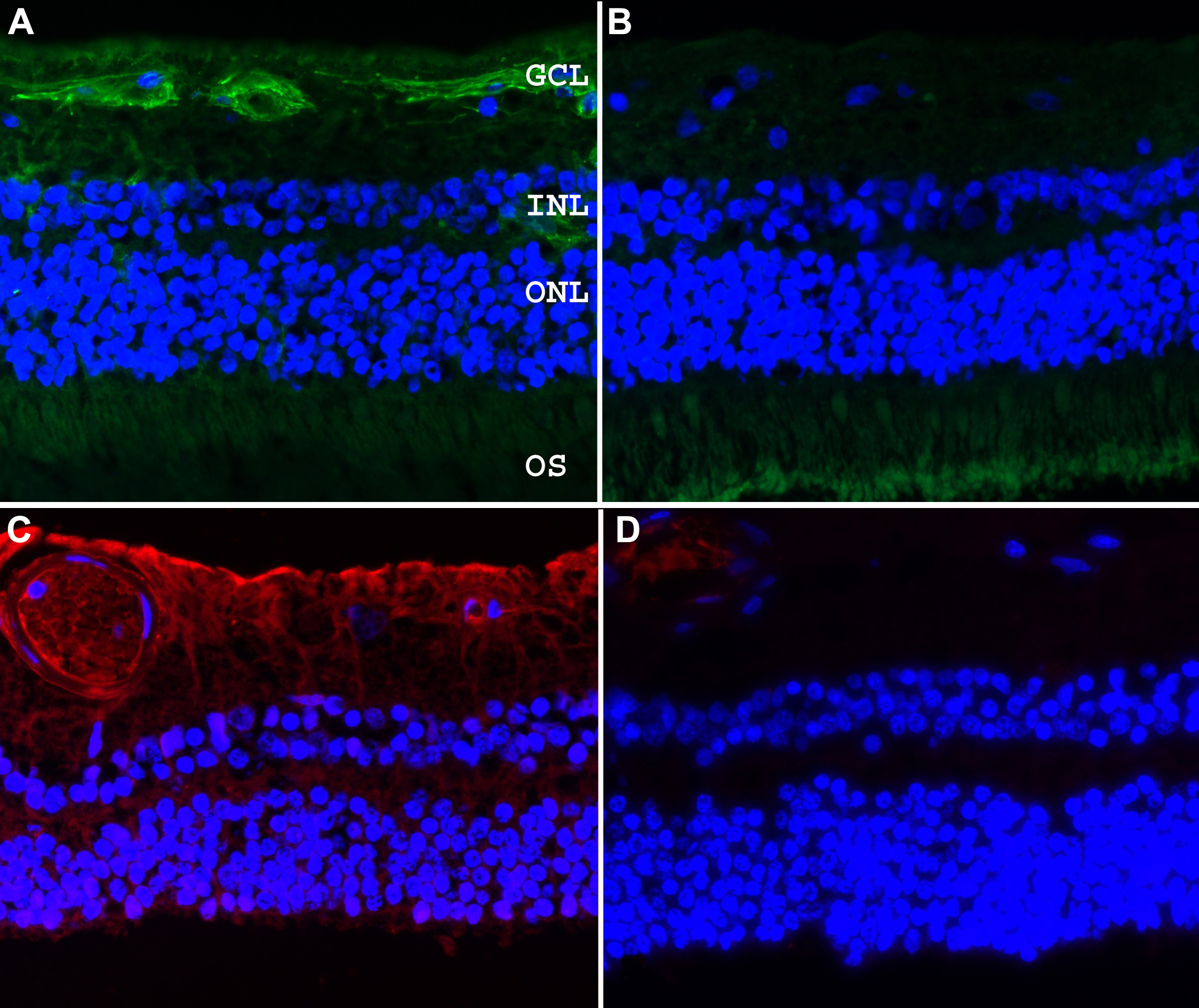
 Figure 1 of Kuehn, Mol Vis 2011; 17:430-435.