Figure 1. Rejection kinetics of corneal
allografts in the BN-PVG strain combination. A: An example of a
well healed corneal allograft approximately day 21. B: Typical
appearance of a rejected PVG allograft, arrow indicates epithelial
rejection line (occasionally observed). C: Rejection kinetics
of the BN-PVG strain combination BN-PVG n=15, BN-BN n=9.
Kaplan–Meier-Survival plot.
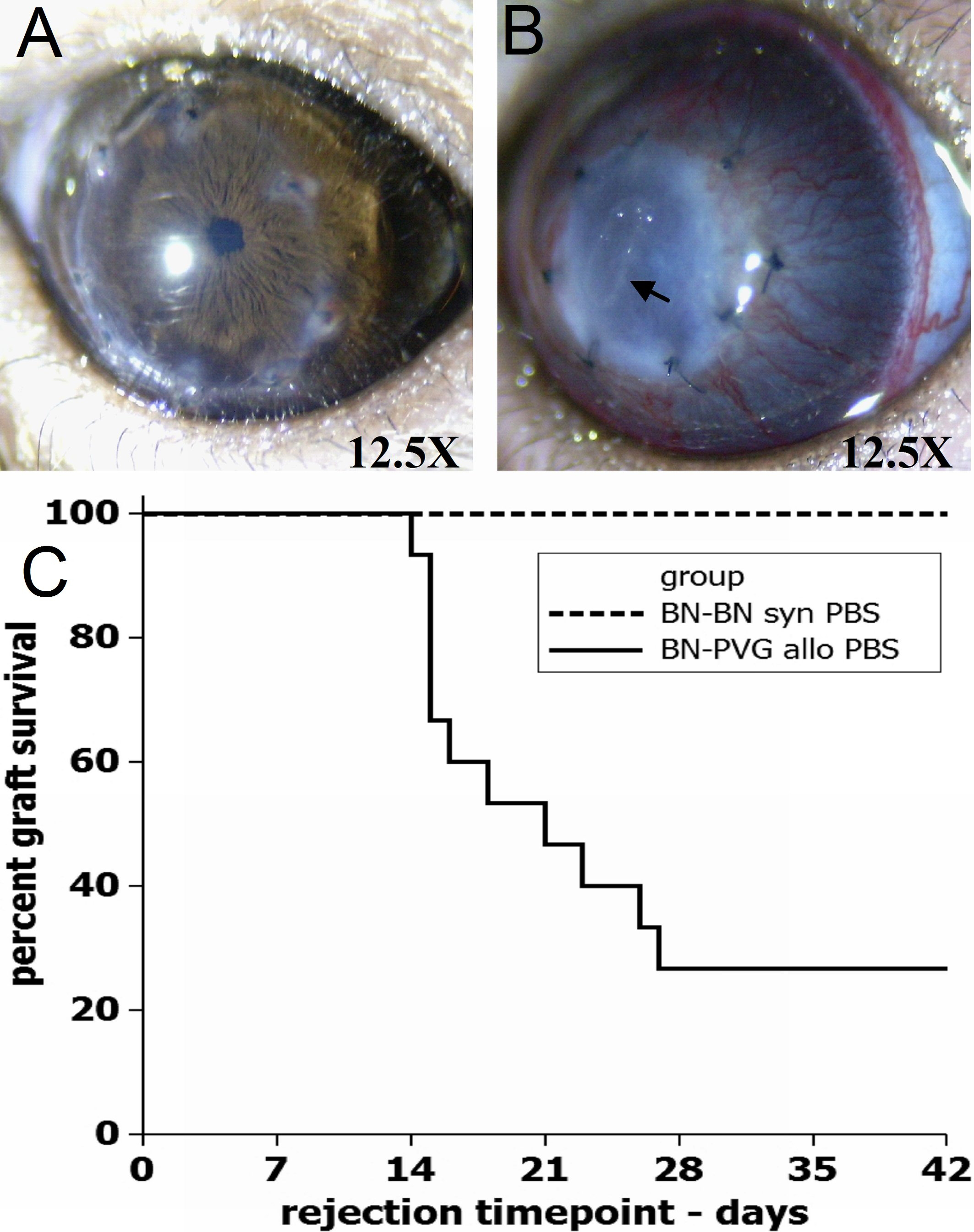
 Figure 1 of Maenz, Mol Vis 2011; 17:420-429.