Figure 3. Light microscopic findings 48 h after alkali burn at 100× magnification. A: The control group and B: amniotic membrane (AM) suspension group. C: Serum eyedrop group and D: comparison group without any manipulation. The figure is representative of the experiments. The corneal thickness was greatly
increased and many polymorphonuclear leukocytes infiltrated into the corneal stroma in the control group (A). However, relatively fewer polymorphonuclear leukocytes infiltrated into the cornea in the AM suspension (B) and serum eyedrop groups (C). Note that the comparison group without any manipulation (D) shows normal corneal thickness without any polymorphonuclear leukocytes infiltration.
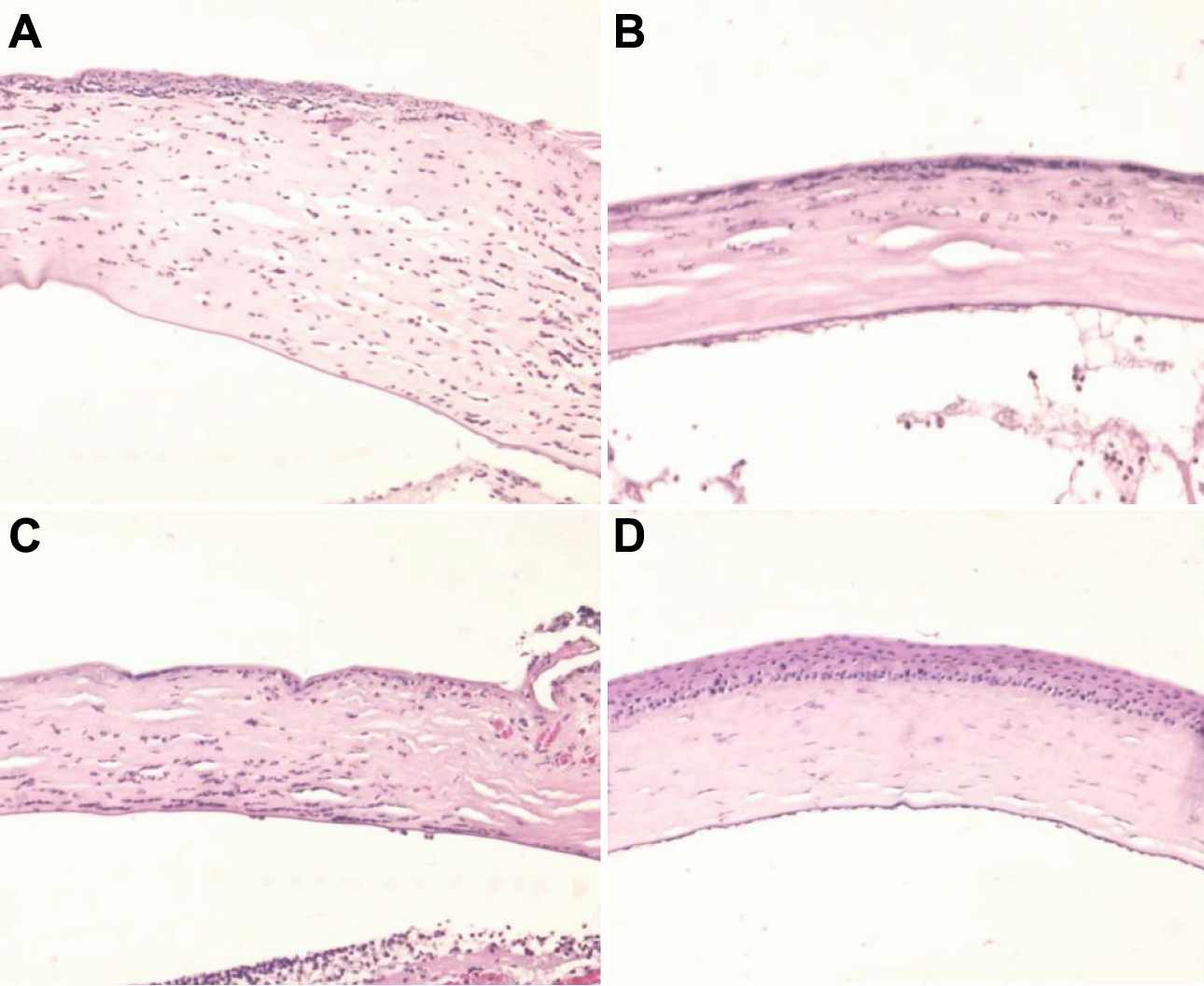
 Figure 3 of
Choi, Mol Vis 2011; 17:404-412.
Figure 3 of
Choi, Mol Vis 2011; 17:404-412.