Figure 4. Increased acetylated histone 3
and acetylated histone 4 levels in retinal ganglion cells after histone
deacetylase inhibitor treatment. A-D: Intensity of
acetylated histone (AcH) 3 and AcH4 expression in retinal ganglion
cells (RGCs) with or without histone deacetylase inhibitor (HDACi)
treatment after 24 h in culture. A, C: In comparison to
controls, sodium butyrate (SB) 0.1 mM significantly increased the
amount of AcH3 (A, *p=0.0119) and AcH4 (C; *p=0.0105) in
RGCs. The expression quotients were 1.7 and 1.4, respectively,
indicating an apparent hyperacetylation after SB treatment. The
horizontal line in A+C indicates, that the difference in
hyperacetylation was statistically significant. B, D:
Valproic acid (VPA) slightly increased AcH3 (B, p=0.1368) and
AcH4 (D, p=0.3326) over baseline levels in controls; however,
differences were not statistically significant (the expression
quotients were 1.2 and 1.1, respectively).
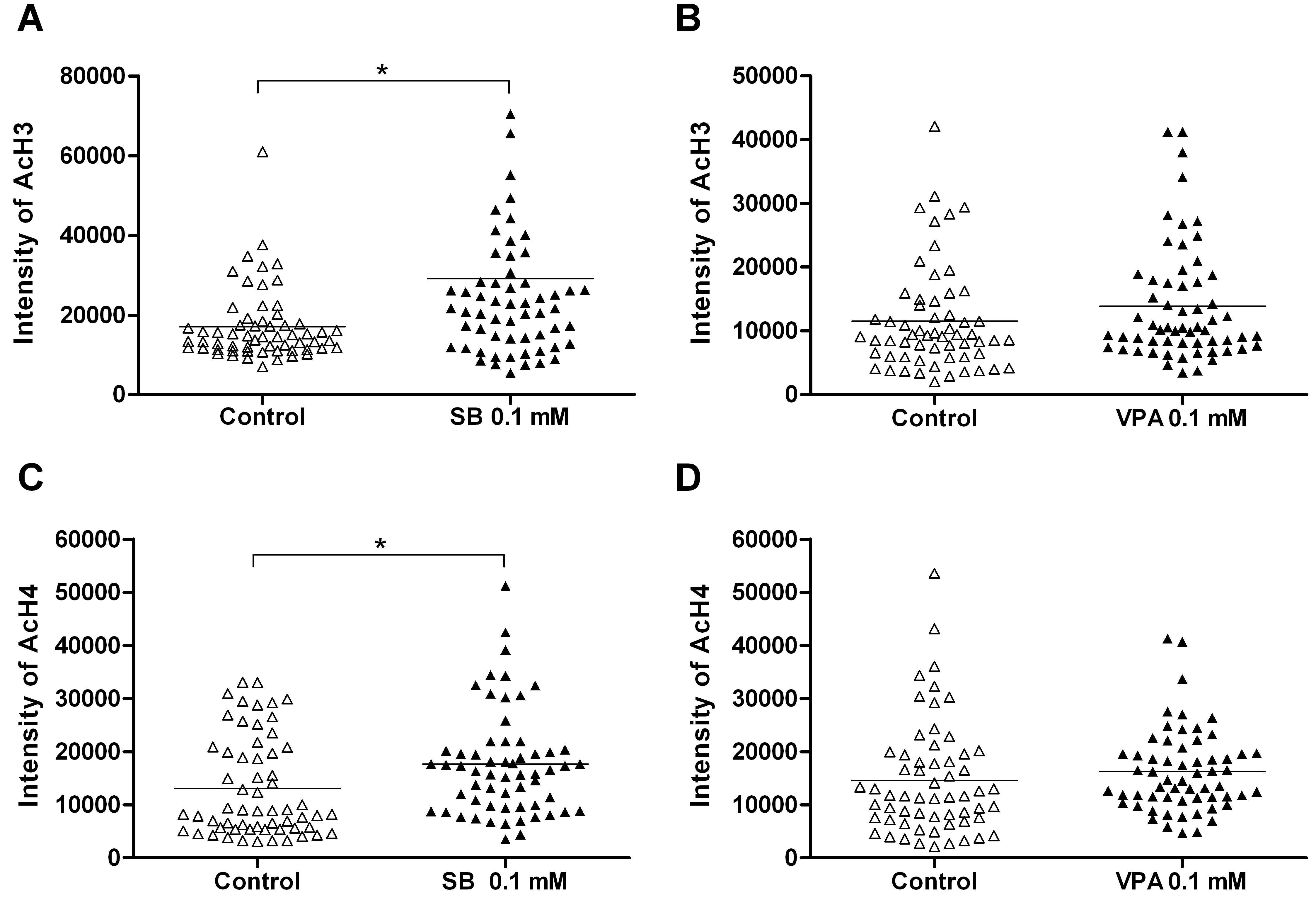
 Figure 4 of Biermann, Mol Vis 2011; 17:395-403.