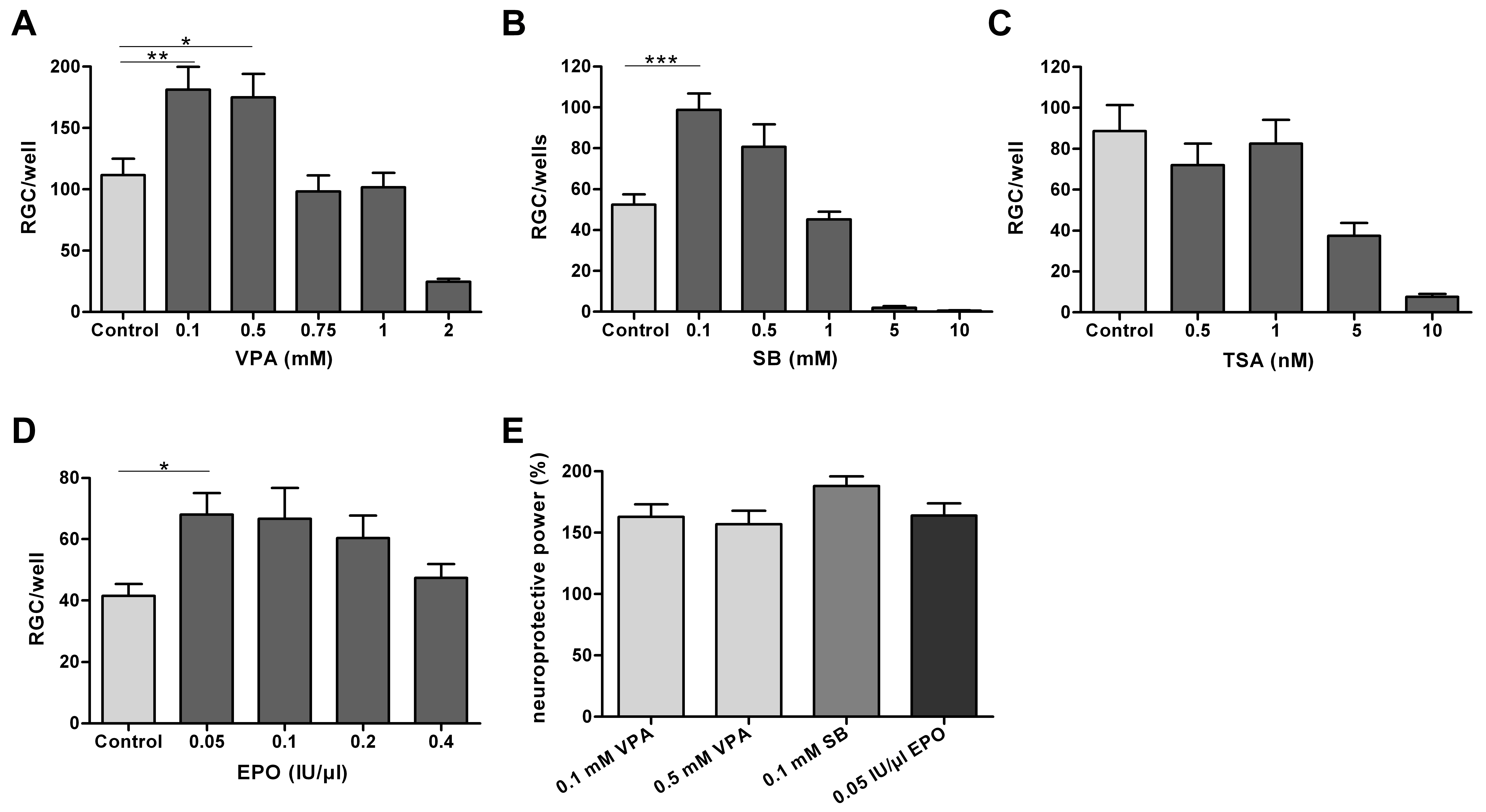
Figure 2. Retinal ganglion cell survival. A-D:
Graphs
showing the dose-dependent effect of histone deacetylase
inhibitors and erythropoietin on retinal ganglion cell (RGC) survival
after 48 h in culture. Data are presented as means and standard error
of the mean (each diagram summarizes four to six culture experiments;
each of the experiments tested the drug in quadruplicates to
hexaplicates of indicated concentrations). A: Compared with
untreated controls, valproic acid (VPA) 0.1 and 0.5 mM significantly
increased RGC survival to 163% (**p<0.01) and 157% (*p<0.05),
respectively. Higher VPA concentrations (0.75 and 1.0 mM) had no
protective effect, while VPA 2 mM significantly decreased RGC survival
to 22% (p<0.001). B: Sodium butyrate (SB) 0.1 mM
significantly increased RGC survival to 188% (***p<0.001), while a
nonsignificant protective effect was seen with SB 0.5 mM. SB 1.0 mM did
not affect RGC survival compared to controls. Higher concentrations of
SB (≥5 mM) significantly decreased RGC survival. C:
Trichostatin A (TSA) 0.5 and 1.0 nM had no effect on RGC survival,
while TSA 5 nM and 10 nM significantly decreased RGC survival to 40%
(p<0.01) and 8% (p<0.001), respectively. D:
Erythropoietin (EPO) 0.05 IU/µl increased RGC survival to 164%
(*p<0.05), while higher concentrations of EPO were less effective. E:
Histone
deacetylase inhibitors (HDACi: VPA 0.1 and 0.5 mM and SB 0.1
mM) had similar protective effects as EPO in this series of experiments
(each column represents the increase in RGC survival [%] after HDACi
treatment compared to individual controls [neuroprotective power=mean
cell numbers treatment/mean cell numbers control*100]).

 Figure 2 of Biermann, Mol Vis 2011; 17:395-403.
  Figure 2 of Biermann, Mol Vis 2011; 17:395-403.