Figure 1. Viable purified retinal ganglion
cells in culture after 48 h. A, B: Representative phase
contrast micrographs showing retinal ganglion cell (RGC) controls (A,
pure
media) and RGCs after treatment with histone deacetylase
inhibitors (HDACi; B, VPA) after 48 h in culture. RGCs (white
arrows) can be discriminated from other retinal cells (black arrows) by
their large cell diameter and fine neurites with branching growth
cones, thus fulfilling the criteria for being counted as viable RGCs.
Larger numbers of degenerated RGCs (asterisk in A) were present
in the control wells. Each scale bar is 50 µm.
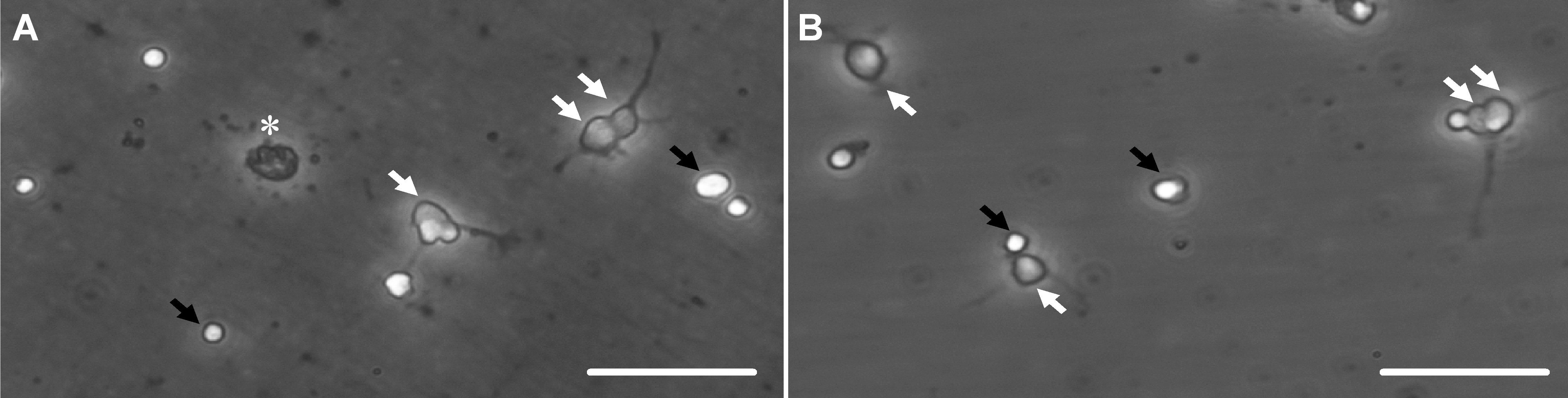
 Figure 1 of Biermann, Mol Vis 2011; 17:395-403.