Figure 1. Effects of curcumin on immunohistochemistry of αA- and αB-crystallin in the eye lens of Wistar rat pups exposed to selenium.
A: This image shows the control lens incubated in saline alone without any antibody treatment. B: This is a control lens that was incubated with saline and subjected to antibody treatment. C: Lens from rat pups administered with selenium alone. D: Lens from rat pups administered with selenium and curcumin simultaneously. E: Lens from rat pups administered with selenium first and then treated with curcumin after 24 h. F: Lens from rat pups pretreated with curcumin and then administered with selenium after 24 h. Lens sections were preincubated
with αA- and αB-crystallin polyclonal antirabbit Immunoglobulin G (IgG) antibody (1:3,000 dilution) and subsequently with
goat anti-rabbit IgG-horse radish peroxidase (HRP) conjugate (1:3,000 dilution). The immunoreactivity was developed with 0.01%
3,3-diaminobenzidine tetrahydrochloride (DAB) and H2O2. Note the brown color formation indicative of peroxidase reaction in the nucleus. Image G, negative control, lens treated with goat anti-rabbit IgG-HRP and developed using DAB and hydrogen peroxide (H2O2). The figure shows the high level of αA- and αB-crystallin expression induced by selenium-mediated oxidative stress. This
increased crystallin expression and aggregate formation was prevented by curcumin pretreatment.
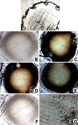
 Figure 1 of
Manikandan, Mol Vis 2011; 17:388-394.
Figure 1 of
Manikandan, Mol Vis 2011; 17:388-394.