Figure 3. Representative images of affected members in the three families were compared by slit-lamp examination and in vivo LSCM. A-D: patient I:2 from family A; E-H: patient II:13 from family B; I-L: patient II:7 from family C. The eye of the proband’s mother revealed grayish annular and crumb-like opacities covered almost
the entire cornea (A). The image of the proband’ sister showed multiple star-like and rounded opacities occupying the central cornea (E). The eye of the proband’s daughter presented with typical snowflake-like opacities combined with linear opacities (I). Focal deposits of reflective material with irregular edges were observed at different levels by LSCM. The images represented
23 μm (B, F, J), 45 μm (C, G, K), and 96 μm (D, H, L) from the corneal surface.
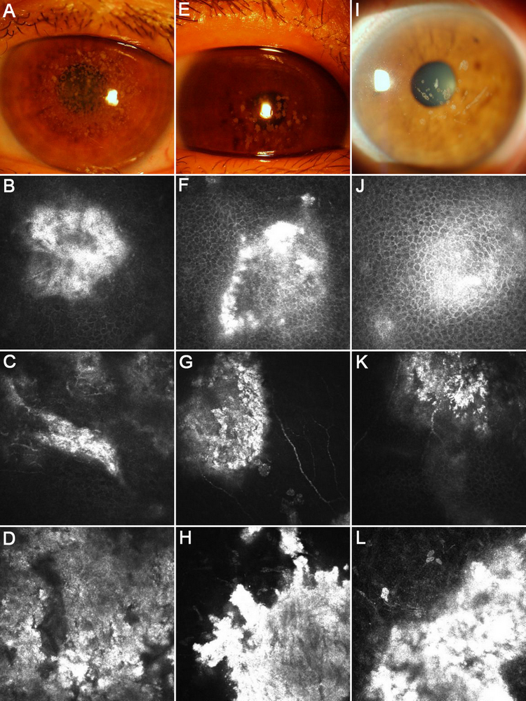
 Figure 3 of
Zhang, Mol Vis 2011; 17:380-387.
Figure 3 of
Zhang, Mol Vis 2011; 17:380-387.