Figure 2. Slit-lamp photomicrographs. In family A, discrete dot opacities in the central cornea of the right eye were noted in the proband
(A). In family B, the right eye of the proband showed a few grayish irregular and rounded corneal opacities (B and C). The peripheral cornea and the cornea between the opacities remained clear. In family C, the image of patient II:5 revealed
scattered annular opacities (D). The proband presented with snowflake-like opacities combined with a linear opacity (E and F).
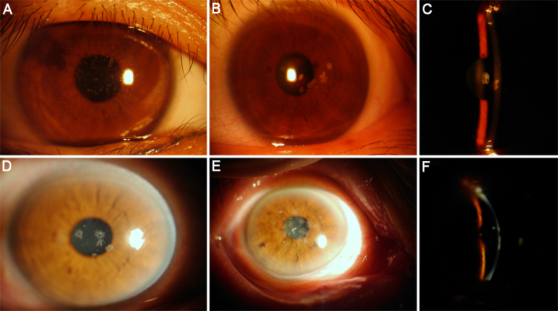
 Figure 2 of
Zhang, Mol Vis 2011; 17:380-387.
Figure 2 of
Zhang, Mol Vis 2011; 17:380-387.