Figure 18. Neuronal processes are often apposed without synaptic contacts. A: A dendrite (purple) from ON ganglion cell C7594 courses through the inner plexiform layer and is physically apposed to many
cells with which it never makes synaptic contact, such as an amacrine cell dendrite (AC, orange). B: An enlarged view of the apposition shown in panel A. The amacrine cell membrane is directly apposed to ganglion cell C7594 with no intervening glial processes (M). The image
is centered on x 71300, y 46622, z 279 in RC1, the apposition spans sections 275–286 (770–990 nm), and is at least 1450 nm
long in the XY plane. C: An apposition between an ON cone bipolar cell (BC, azure) and ganglion cell C7594 at a more distal location in RC1. E denotes
glutamate; γ denotes GABA; τ denotes taurine. The letter colors match the profiles in the image. The bipolar cell makes a
ribbon monad onto a very small amacrine cell dendrite, but is never presynaptic to the ganglion cell. The image is centered
on x 73722, y 53700, z 258 in RC1, spans sections 256–269 (910–1170 nm), and is at least 2200 nm long in the XY plane based
on serial tracking. Scales, 1000 nm.
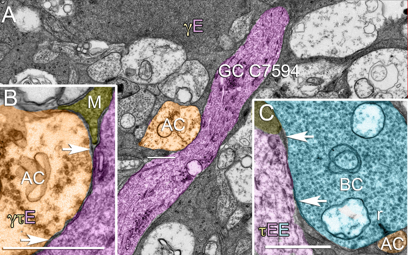
 Figure 18 of
Anderson, Mol Vis 2011; 17:355-379.
Figure 18 of
Anderson, Mol Vis 2011; 17:355-379.