Figure 12. Axonal ribbon synapses (black circles) and veto synapses (white circles) are distributed along the axons of 105 reconstructed
bipolar cells. Grey lines are OFF cone bipolar cells terminating high in the inner plexiform layer. Green lines are G+ ON
cone bipolar cells. Magenta lines are rod bipolar cells with extensive AI and AII amacrine cell contacts. Each line indicates
the length of the axon from its point of entry to its terminal expansion level in the inner plexiform layer (IPL). The physical
cells are longer as we show only the axon, not the entire terminal arbor. The break in the lower panel axons represents the
approximate position of the lower limit of identified OFF bipolar cell processes. Importantly, some bona fide ON bipolar cells
axons are shorter than the longest OFF bipolar cell axons and terminals and they co-mingle.
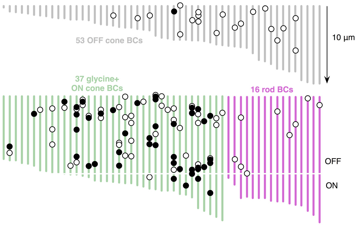
 Figure 12 of
Anderson, Mol Vis 2011; 17:355-379.
Figure 12 of
Anderson, Mol Vis 2011; 17:355-379.