Figure 11. RC1 contains novel retinal
networks. A: A Viking rendering of γ+ amacrine cell C5303 shows
that it is postsynaptic to ON cone bipolar cells at axonal ribbon sites
(circles C and D), presynaptic to ON cone bipolar cell C483, and
co-stratifies with ON starburst amacrine cell C4890 (scale 20 μm). The
circles indicate corresponding transmission electron microscope (TEM)
images. B: Axonal ribbons (r) from ON cone BC C180 target AC
neurites as the axon bifurcates in mid-inner plexiform layer. C:
ON
cone BC C177 makes axonal nanoribbon contacts onto γ+ amacrine cell
C5303. D: Axonal ribbon synapses from C166 target cell C5303.
C166 receives an axonal veto synapse from a yet unidentified amacrine
cell (arrow). E: A large γ+ amacrine cell makes an axonal veto
synapse onto an OFF cone bipolar cell axon. F: An axonal
cistern contact is formed between ON cone bipolar cell C168 onto an
amacrine cell process. E denotes glutamate; γ denotes GABA; and
question mark denotes unknown. The letter colors match the profiles in
the image. The scales for images B-F are 500 nm.
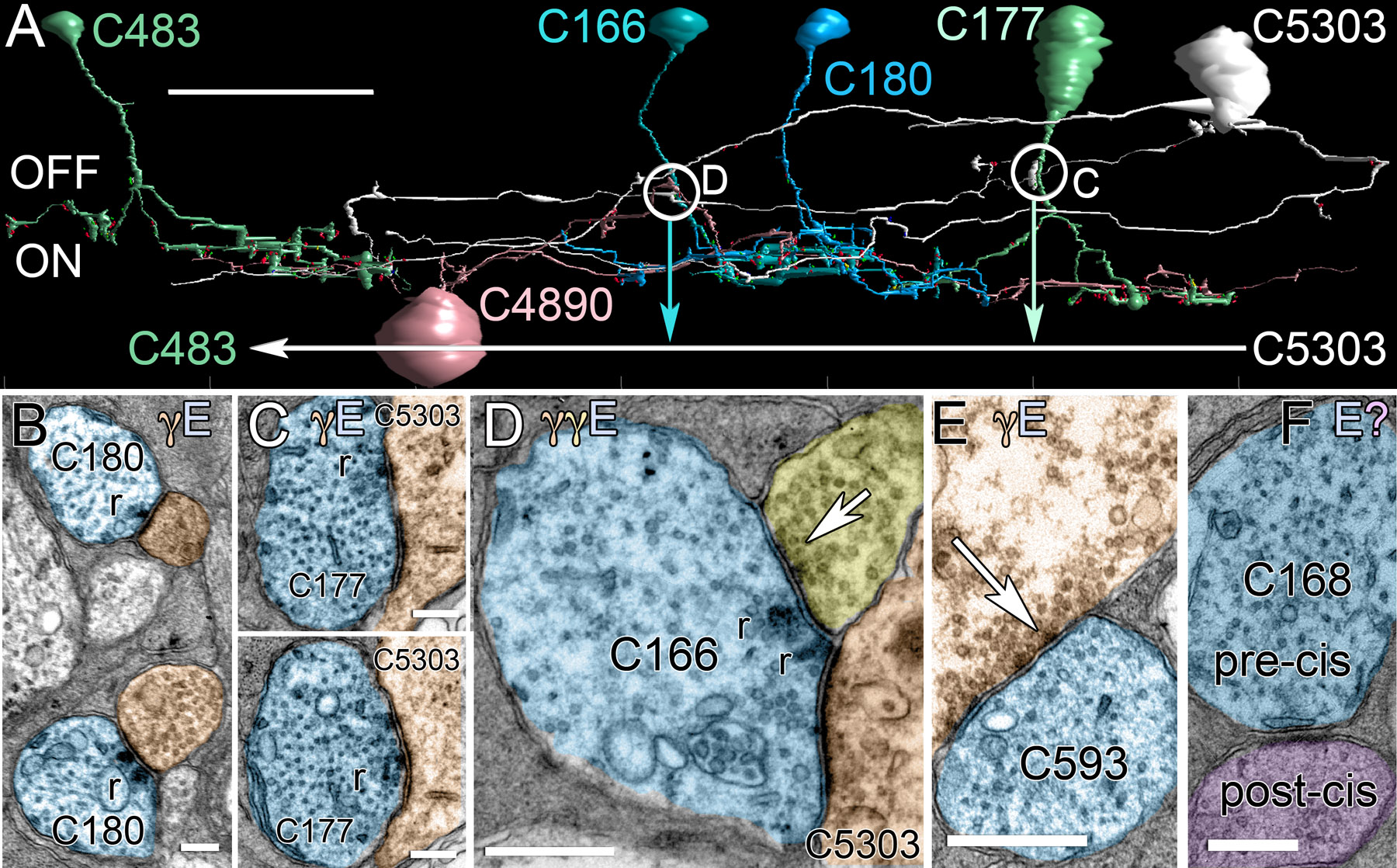
 Figure 11 of Anderson, Mol Vis 2011; 17:355-379.