Figure 6. Epithelial cells obtained from full thickness and superficial limbal explants cultured for 3 weeks in cholera toxin-free Green
medium. Staining for CK3 (A: superficial limbal explants; B: full thickness limbal explants), vimentin (C: superficial limbal explants. D: full thickness limbal explants), broad spectrum cytokeratins (CK4, 5, 6, 8, 10, 13, and 18; E: superficial limbal explants; F: full thickness limbal explants), CK19 (G: superficial limbal explants; H: full thickness limbal explants), delta N p63α (I: superficial limbal explants; J: full thickness limbal explants), ABCG2 (K: superficial limbal explants; L: full thickness limbal explants) of limbal cells; M: negative control; N: positive control (human corneal epithelial cells stained with CK3). O: keratocytes stained with vimentin. Bars: 200 µm; magnification: 10×.
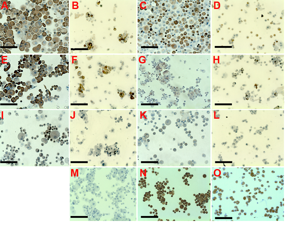
 Figure 6 of
Ghoubay-Benallaoua, Mol Vis 2011; 17:341-354.
Figure 6 of
Ghoubay-Benallaoua, Mol Vis 2011; 17:341-354.