Figure 5. Immunohistochemical staining of normal central cornea and limbus. Vimentin (A, B), delta N p63α (C, D), cytokeratin 3 (E, F), cytokeratin 19 (G, H), and broad spectrum cytokeratins (MNF116; I, J) in normal central cornea and limbus. Vimentin is not detected in corneal epithelial cells (A) but is detected in the basal limbal epithelial layer and stromal cells (B). Delta N p63α is strongly expressed in the basal layer of the limbus (D). The entire superficial limbal epithelial layer and cells in the mid layer are positive for CK3 (F). CK19 labels the basal and superficial limbal epithelial layers whereas cells in the mid layer are not stained by anti-CK19.
Bars: 200 µm; magnification: 10×.
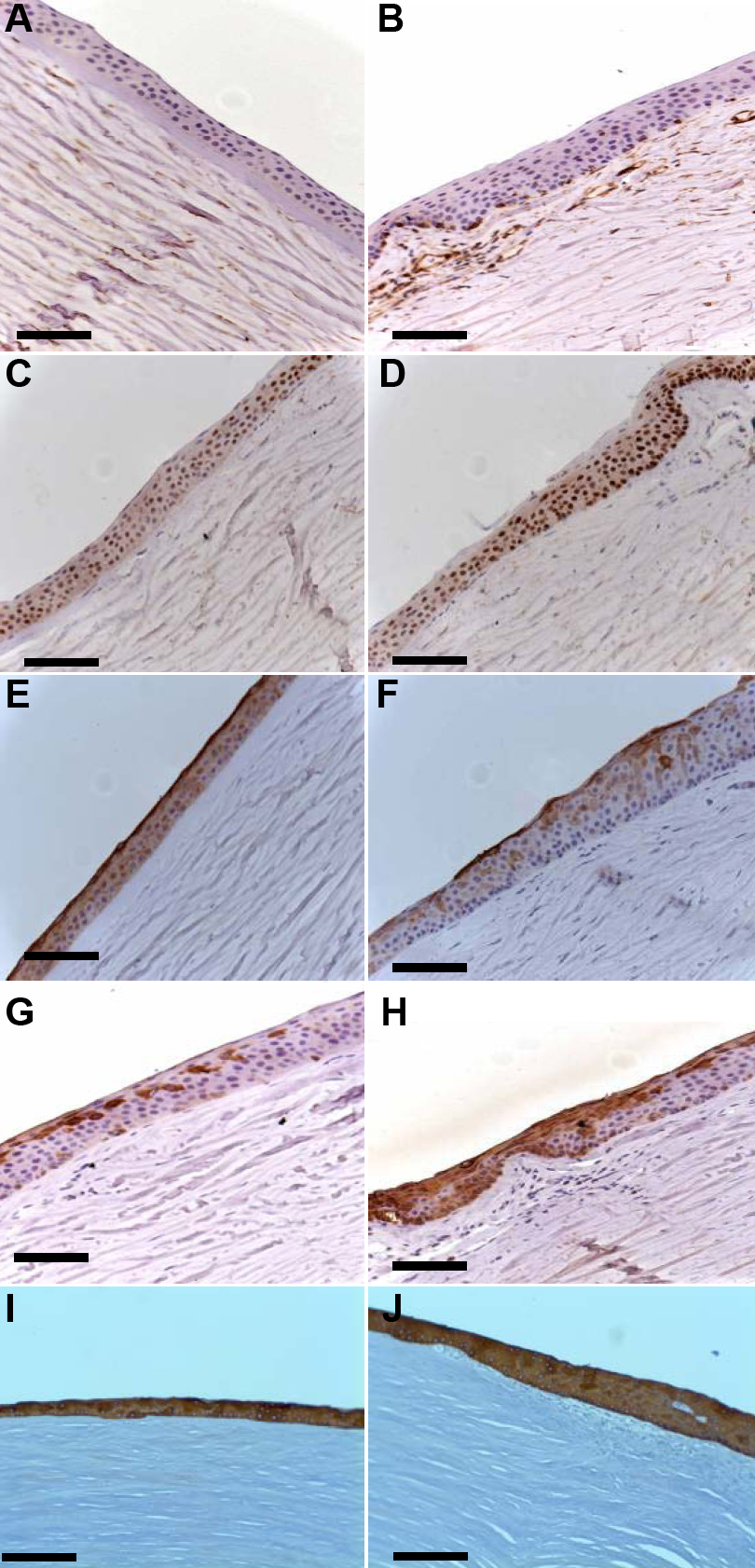
 Figure 5 of
Ghoubay-Benallaoua, Mol Vis 2011; 17:341-354.
Figure 5 of
Ghoubay-Benallaoua, Mol Vis 2011; 17:341-354.