Figure 1. Limbal epithelial cells cultured from human explants. A: Superficial limbal explant with polygonal cells covering the well after three weeks. B: Polygonal and fibroblast-like cells from full-thickness limbal explant. C: Fibroblast-like cells from stromal explant.
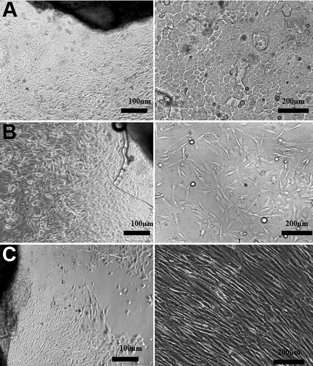
 Figure 1 of
Ghoubay-Benallaoua, Mol Vis 2011; 17:341-354.
Figure 1 of
Ghoubay-Benallaoua, Mol Vis 2011; 17:341-354.