Figure 1. Schematic picture shows the different stages of pterygia. Stage 1: The head of pterygia did not reach the midline between
the limbus and pupillary margin. Stage 2: The head of pterygia passed the midline but did not reach the pupil. Stage 3: The
head of pterygia passed the pupillary margin.
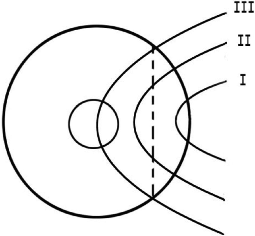
 Figure 1 of
Chao, Mol Vis 2011; 17:23-31.
Figure 1 of
Chao, Mol Vis 2011; 17:23-31.