Figure 5. Quantification of the
retinal area with neovascularization in P17 normal air mice and
P17 oxygen-induced retinopathy (OIR) mice. The P17 normal air
mice (control; A) and P17 OIR mice were given a
retro-orbital injection (B) or left-ventricular perfusion
(C) of fluorescein isothiocyanate dextran (FITC)-dextran.
The neovascularization areas were outlined with Image Pro-Plus
5.1 software (Media Cybernetics Company, Silver Spring, MD). The
percentage of the retinal neovascularization in the OIR group is
a significant increase when compared to the control group (n=6),
and no statistical difference was found between the
retro-orbital injection (RO) group (n=6) and the LV group (n=6)
D: Data shown are mean±standard deviation, *p<0.05.
NV: neovascularization, LV, left-ventricular perfusion. Scale
bar: 100 μm.
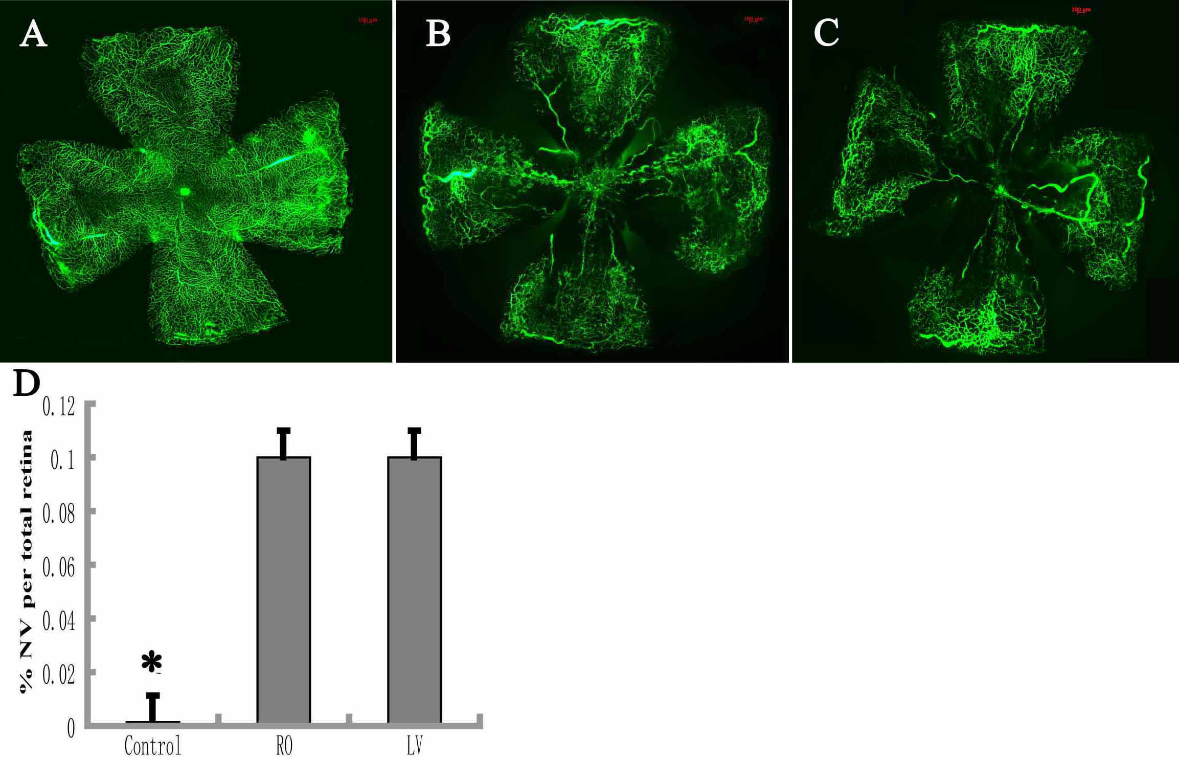
 Figure 5
of Li, Mol Vis 2011; 17:3566-3573.
Figure 5
of Li, Mol Vis 2011; 17:3566-3573.