Figure 2. The superficial and deep
vascular plexuses of the retina in a P17 oxygen-induced
retinopathy mouse after a retro-orbital injection of fluorescein
isothiocyanate dextran (FITC)-dextran. A: The retinal
flatmount of a P17 oxygen-induced retinopathy mouse shows the
fluorescence of small retinal vessels, avascular areas, and
neovascular tufts. B: Original magnification: 50×. The
superficial neovascular tufts (Arrow, C) and the deep
vascular plexus (D) can be observed. Original
magnification: 100×.
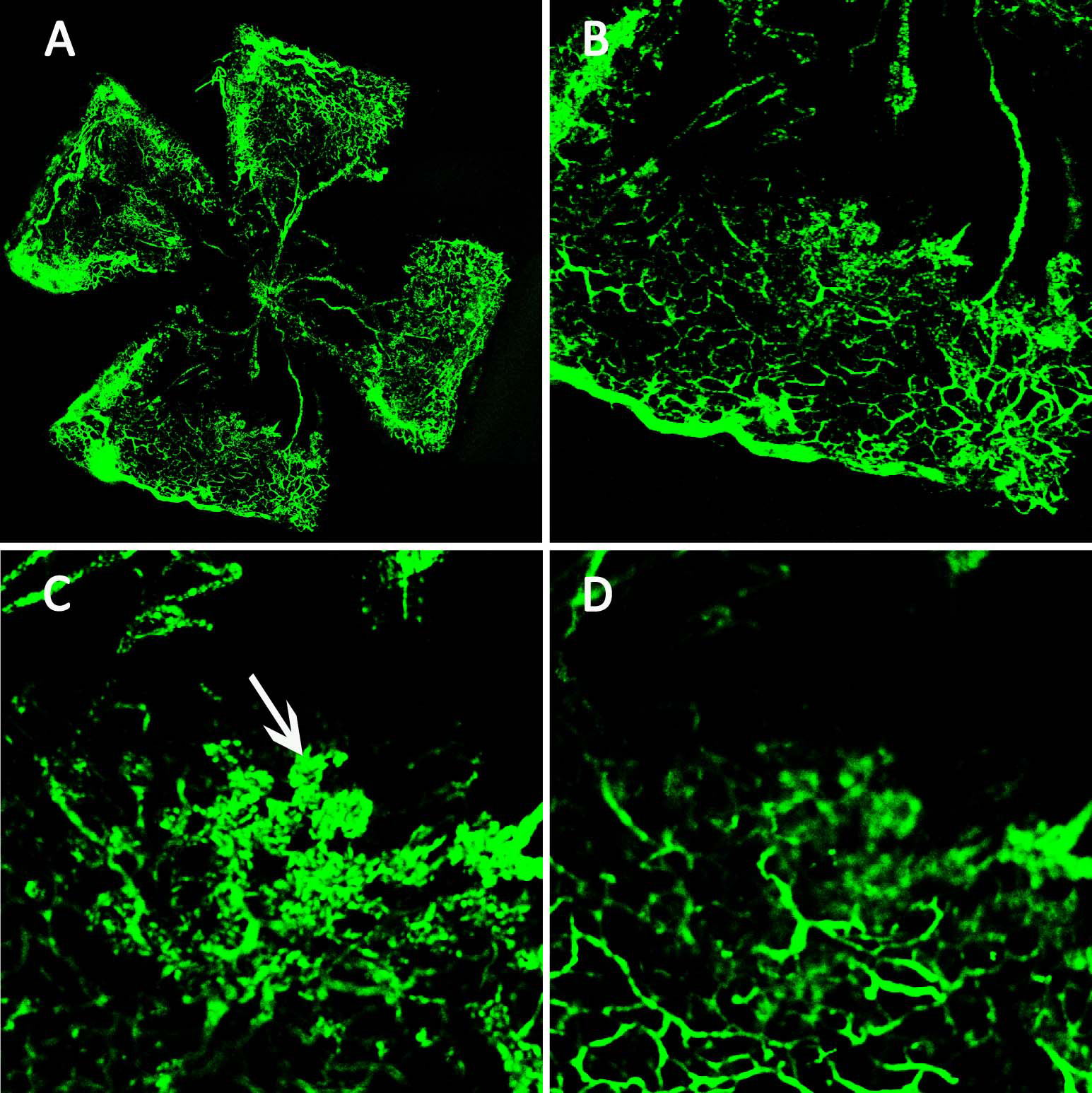
 Figure 2
of Li, Mol Vis 2011; 17:3566-3573.
Figure 2
of Li, Mol Vis 2011; 17:3566-3573.