Figure 4. Unoprostone and M1
suppressed phagocytotic dysfunction induced by white light in
ARPE-19. A: Representative fluorescence microscopy shows
latex beads and morphology, or latex beads and nuclear staining
for Hoechst 33342 after 48 h light irradiation. Arrowheads
indicate the fluorescent beads. B, C:
Unoprostone (0.001–1 µM), M1 (0.001–1 µM), and PGF2α
(1 µM) or latanoprost (1 µM) were added before light
irradiation. Latex beads were added after light irradiation, and
4 h later, they were removed by washing. The number of
intracellular latex beads was counted, and phagocytotic activity
was expressed as the percentage of latex beads to total cell
numbers. Data are expressed as mean±SEM (PGF2α and
Lat; n=4, unoprostone and M1; n=8). ** p<0.01 versus vehicle;
## p<0.01 versus control (Dunnett’s test). Con:
control; Veh: vehicle; Lat: latanoprost. Scale bar represents 50
µm.
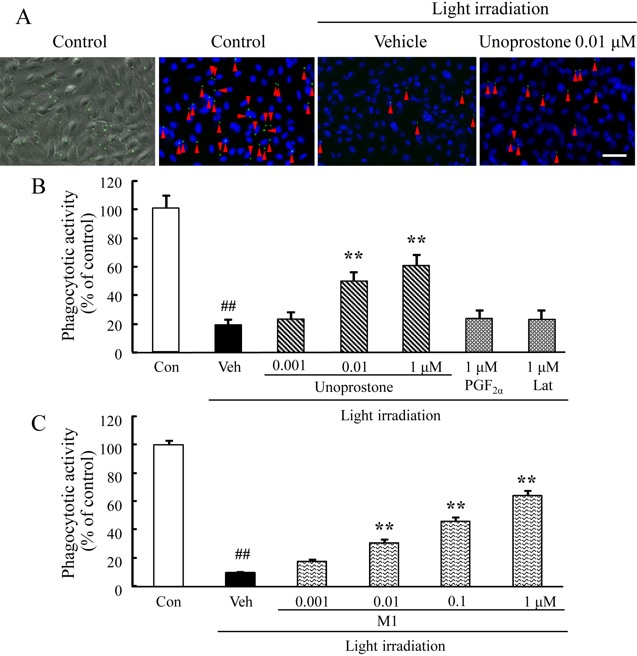
 Figure 4
of Tsuruma, Mol Vis 2011; 17:3556-3565.
Figure 4
of Tsuruma, Mol Vis 2011; 17:3556-3565.