Figure 3. Unoprostone reduced
morphological change induced by light irradiation in mouse
retinal cone-cell line 661W cells. A: Representative
images show cell morphologies. Control cells had an elongated
appearance with extended processes, whereas cell morphology was
altered to a round shape after 24 h light irradiation. Cells
were treated with 10 µM of unoprostone and 100 µM of trolox
before light irradiation. B: Unoprostone (1–10 µM) was
added before light irradiation. The number of nonround-shaped
cells was counted, and expressed as the percentage of total cell
numbers. C: Representative images show JC-1 stained
cells. Healthy cells with mainly JC-1 J-aggregates can be
detected as red cells, and unhealthy or apoptotic cells with
mainly JC-1 monomers can be detected as green cells. Merged
(orange) cells were determined to be pre-apoptotic (early or
middle state of transition to cell death) cells. D: The
number of cells with each color were counted and expressed as
the percentage of total cell numbers (red bar=red fluorescence
cells, orange bar=merged cells, and green bar=green fluorescence
cells, respectively). Data are expressed as mean±SEM (B;
n=6, D; n=3). * p<0.05, ** p<0.01 versus vehicle;
## p<0.01 versus control (Dunnett’s test). Con: control; Veh:
vehicle; Tro: trolox. Scale bar represents 100 µm.
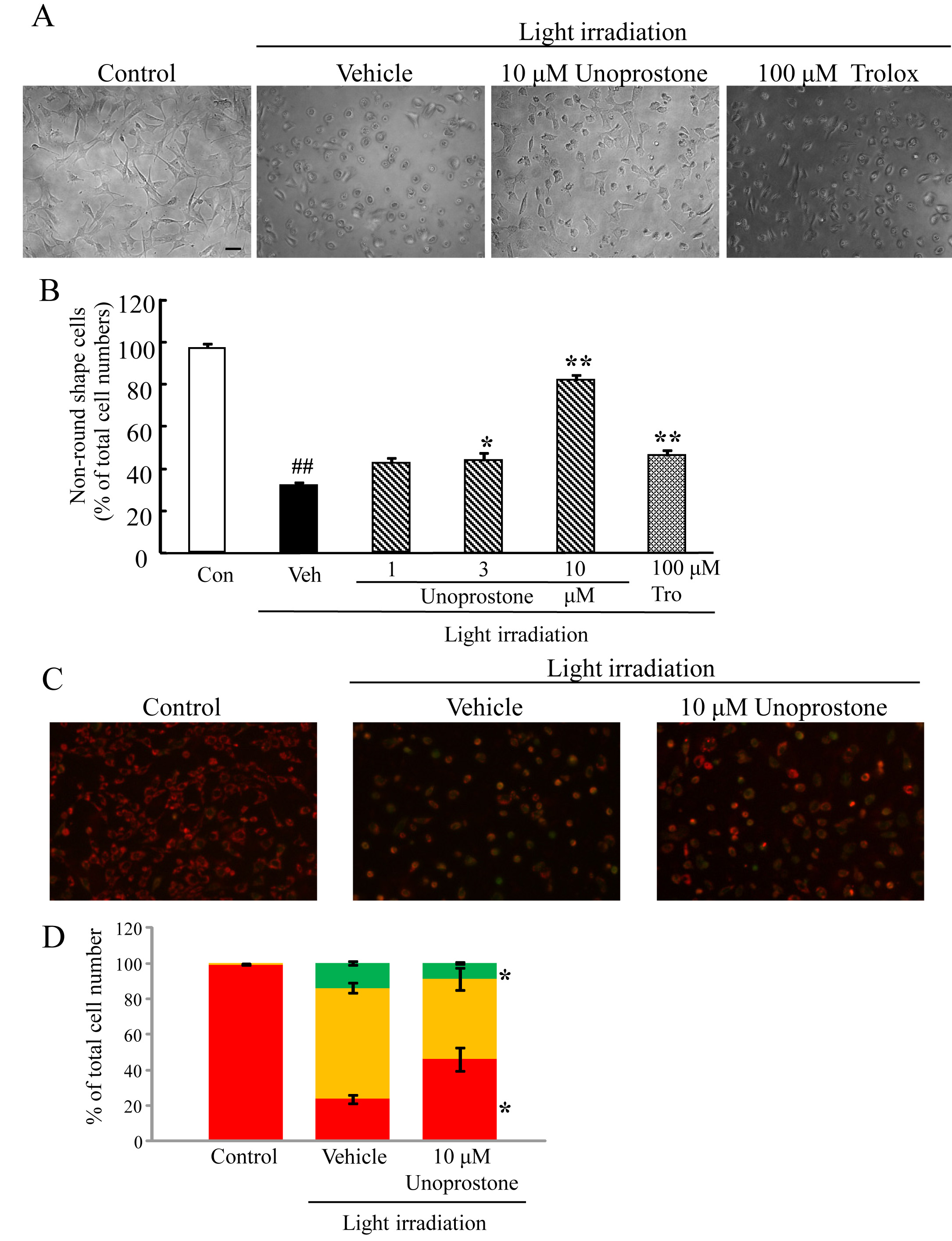
 Figure 3
of Tsuruma, Mol Vis 2011; 17:3556-3565.
Figure 3
of Tsuruma, Mol Vis 2011; 17:3556-3565.