Figure 3. Fundus photographs of
affected individuals from family TB109. Fundus photographs of
individual II-1 at the age of 8 (A), and individual II-2
at the age of 6 (B), demonstrating extensive retinal
degeneration, salt and pepper–like, retinal pigment epithelial
atrophy, reduced foveal reflex, irregularity of vitreoretinal
interface, mild vascular attenuation, and absence of peripheral
bone spicules or pigment deposits. The optic disc looks normal
with no definitive pallor or signs of atrophy.
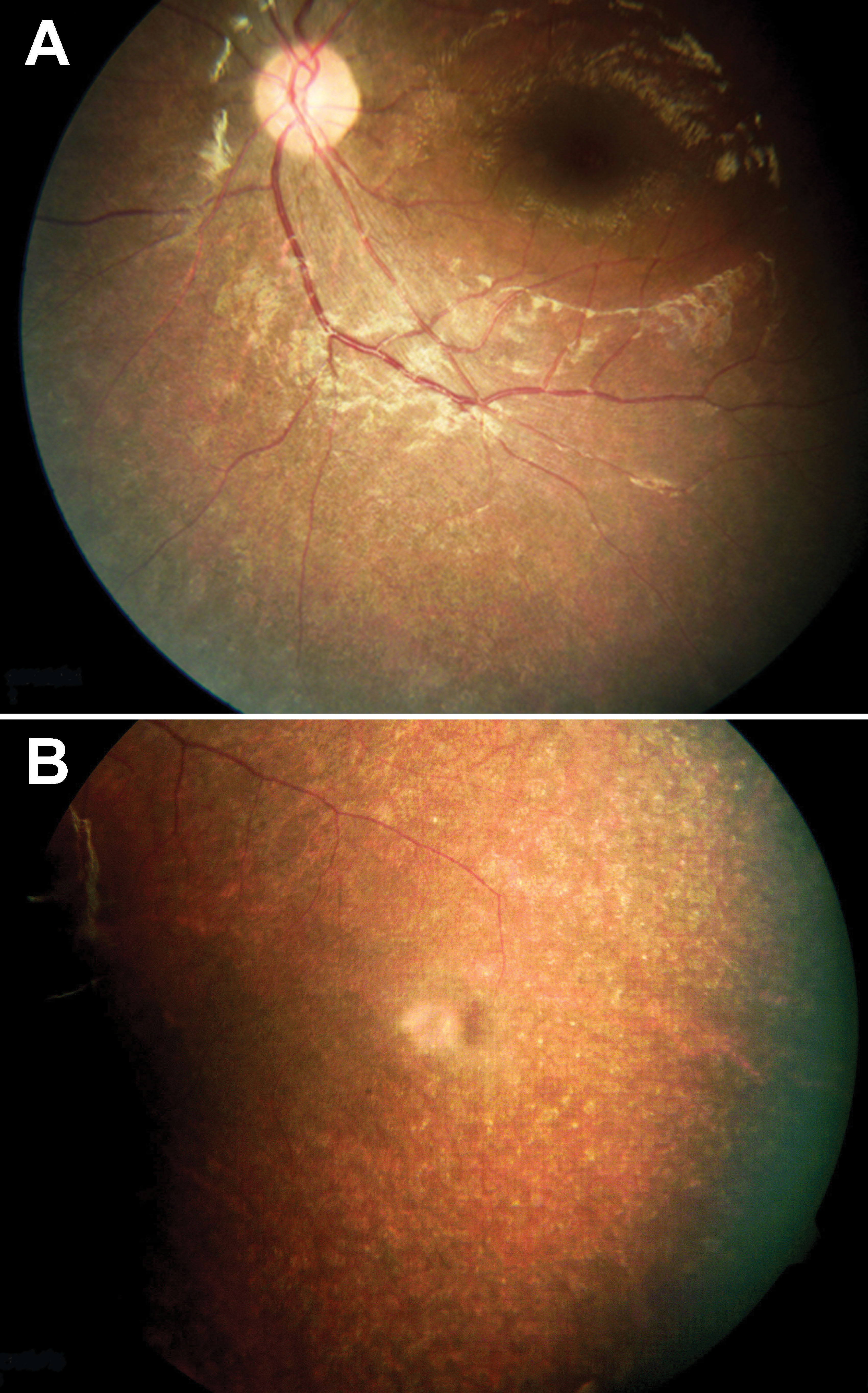
 Figure 3
of Rizel, Mol Vis 2011; 17:3548-3555.
Figure 3
of Rizel, Mol Vis 2011; 17:3548-3555.