Figure 1. Comparison of SDS–PAGE gel
patterns of proteins in camel tear fluids between summer and winter. A:
Proteins
of camel tears in the summer (lane Cs) and in the winter (lane
Cw) were separated on a 13% gel with equal amount of total tear
proteins in each sample. Thirteen well resolved bands are detected in
both lanes. B: Graphic of lane comparison of camel tear
proteins between the summer (dotted line) and the winter (solid line).
B1, Band1; B2, Band2; B3, Band3; and so forth in B are
correspondent with those in A.
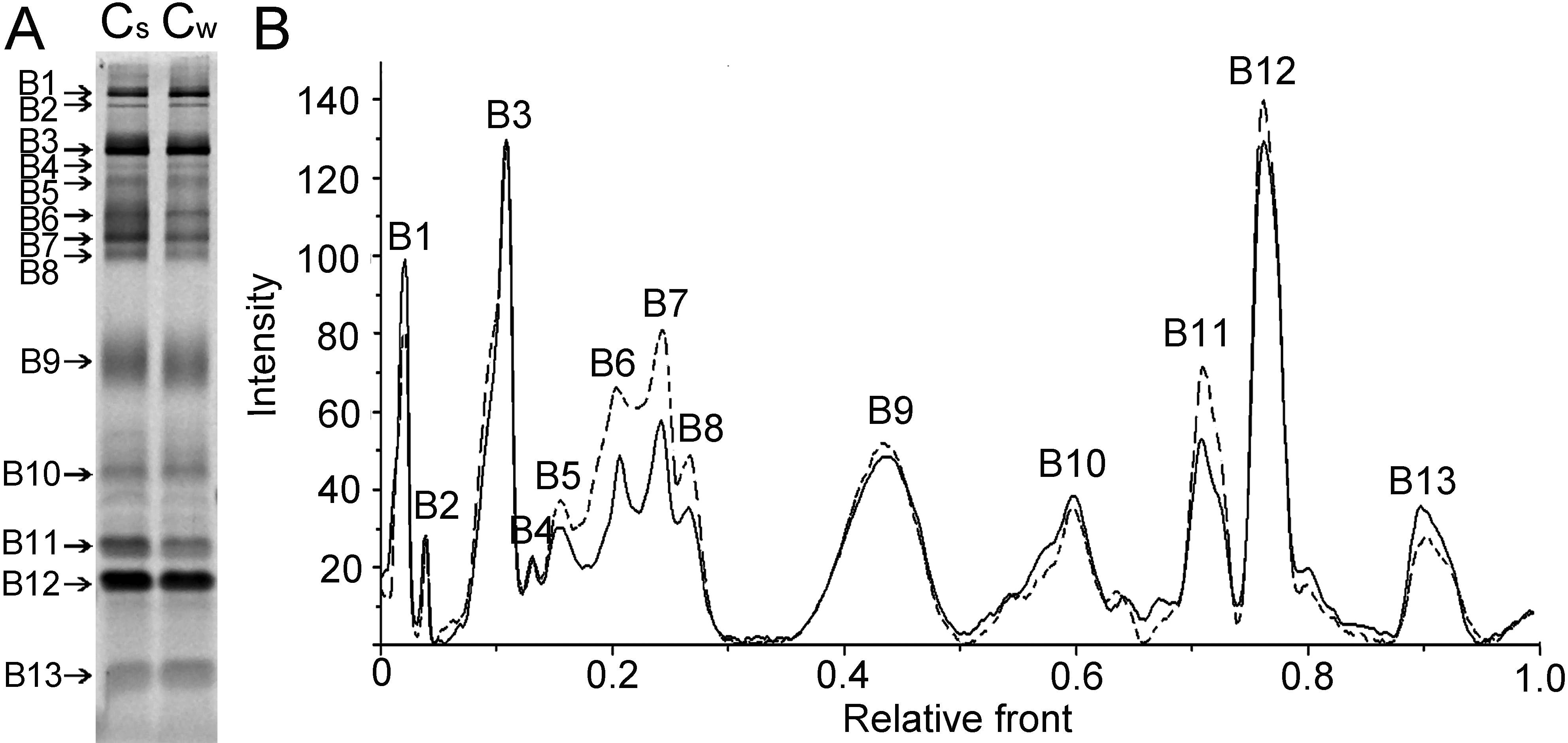
 Figure 1 of Chen, Mol Vis 2011; 17:323-331.