Figure 3. Gene structure of human BBS4.
A: Exon-intron structure of BBS4. Exons are
indicated as black boxes. The single missense mutation
identified is located in the fifth exon (p.E85Q, red arrow). B:
Sequence traces of wild-type and affected family members. A
homozygous mutation from G to C was identified in affected
member KKESH205#7 (red arrow). C: Amino acid alignment
of a portion of the predicted BBS4 protein from nine different
vertebrate species. The mutated amino acid is indicated (p.E85Q,
red arrow).
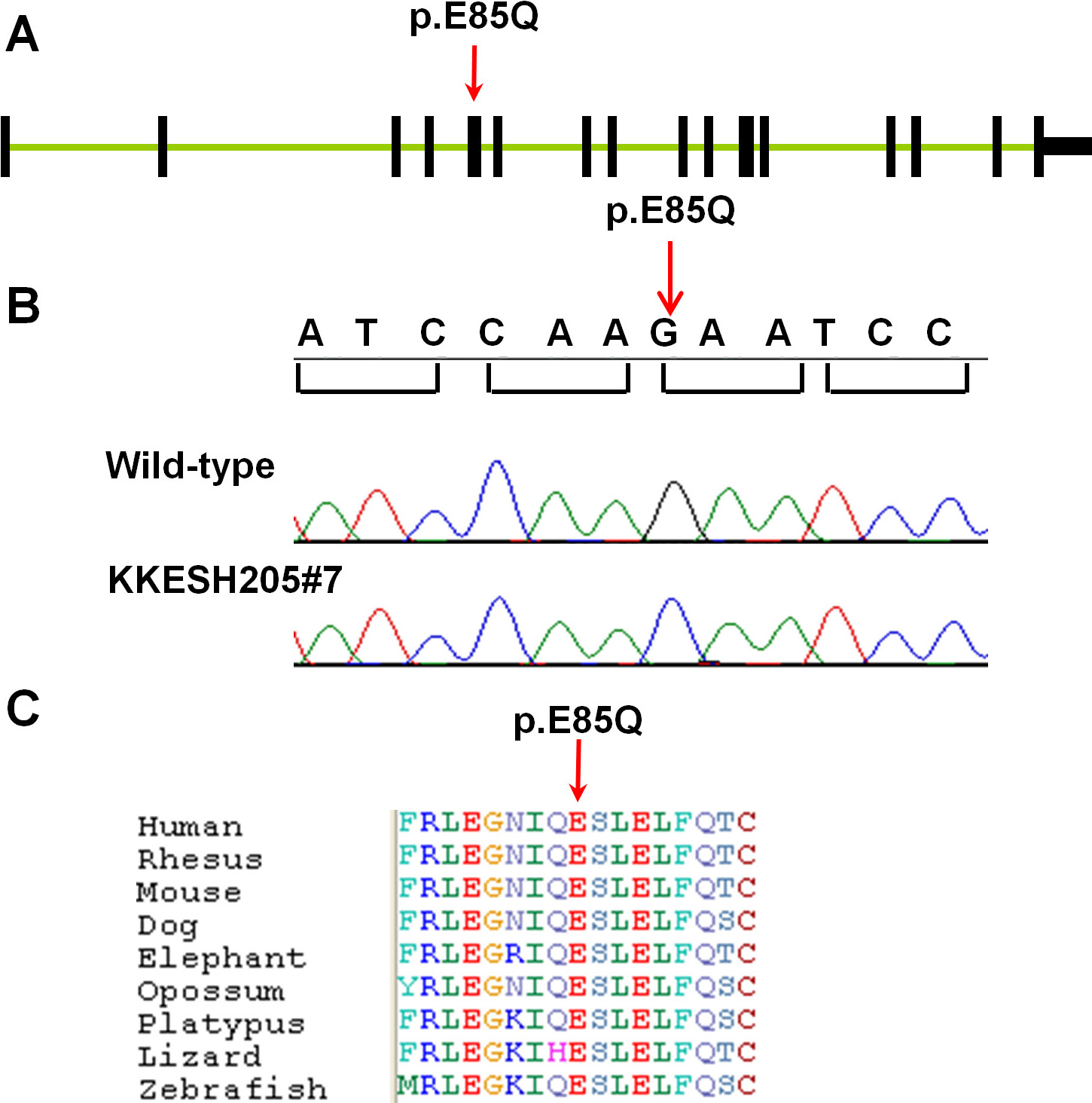
 Figure 3
of Wang, Mol Vis 2011; 17:3529-3540.
Figure 3
of Wang, Mol Vis 2011; 17:3529-3540.