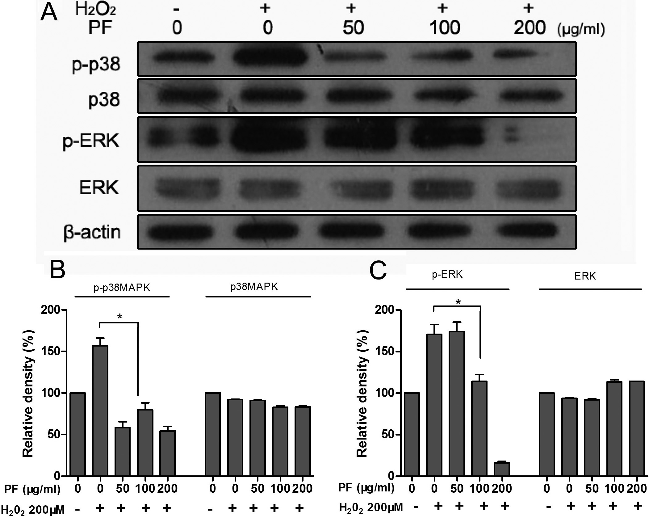
Figure 7. Paeoniflorin (PF) inhibits
H2O2-induced MAPK activation in ARPE-19
cells. ARPE-19 cells were pretreated with different
concentrations of PF for 4 h and exposed to 200 μM H2O2
for 30 min; cell lysates were then prepared. The phosphorylated
and total protein levels of p38MAPK and ERK1/2 were detected
with specific antibodies, using western blot analysis as
described in the Methods. β-actin served as the loading control.
Figures were selected as representative data from three
independent experiments. Quantitative analysis was performed by
measuring the intensity relative to the control. Each value
represents the mean±SEM of three independent experiments (n=3
experiments,*p<0.05).

 Figure 7
of Wankun, Mol Vis 2011; 17:3512-3522.
  Figure 7
of Wankun, Mol Vis 2011; 17:3512-3522.